Cross Section Of a Tooth

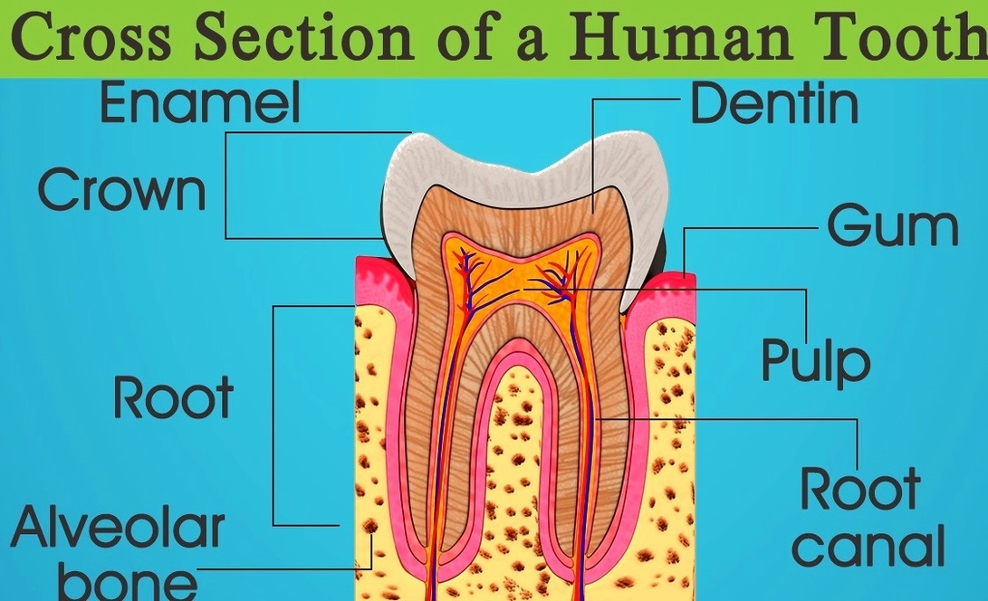
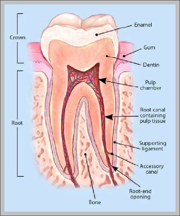
cross section of a molar [1] Teeth are formed of two main parts: the crown (the visible protruding part) and one or several roots (the part inserted into the maxilla). Highly mineralized tissue covering and protecting the dentin of the crown; it is the hardest tissue in the organism.

cross section of a molar Teeth are formed of two main parts: the crown (the visible protruding part) and one or several roots (the part inserted into the maxilla).

Highly mineralized tissue covering and protecting the dentin of the crown; it is the hardest tissue in the organism. Part of the tooth covered with enamel and protruding outside the gum. Narrow part of the tooth surrounded by the gum separating the crown from the root.

Cross Section Of a Tooth Diagram - Chart - diagrams and charts with labels. This diagram depicts Cross Section Of a Tooth

Cross section of a tooth

Picture Of Human Teeth

The human teeth is composed of 16 upper teeth and 16 lower teeth. They are also divided into four quadrants. This article explains the teeth number and names used in the US and Mexico.

Humans have the following types of teeth: 1 Incisors. Incisors are the sharp teeth at the front of the mouth that bite into food and cut it into smaller pieces. They are flat with a thin edge. 2 Canines. 3 Premolars. 4 Molars.

The human teeth dental chart illustrates the location and roles each tooth plays in performing their jobs of cutting, grinding and crushing food. Teeth differ in size, shape and their placement in the jaws.

Picture Of Human Teeth Diagram - Chart - diagrams and charts with labels. This diagram depicts Picture Of Human Teeth

picture of human teeth

Tooth Anatomy

Each part of a tooth has unique functions and properties. Aetna’s Simple Steps to Better Dental Health lists major parts of tooth anatomy, including enamel, dentin, cementum, root (s) and the root canal chamber (s) inside the tooth. Damaged teeth, especially teeth with cracked or eroded enamel, are very susceptible to cavities.

Dental anatomy. Usually, there are 20 primary (“baby”) teeth and 28 to 32 permanent teeth, the last four being third molars or ” wisdom teeth “, each of which may or may not grow in. Among primary teeth, 10 usually are found in the maxilla (upper jaw) and the other 10 in the mandible (lower jaw).

The periodontium consists of: Gingiva: it is a part of the oral mucosa that surrounds teeth and covers the maxilla and mandible. The beautiful smile comes from a harmonious coexistence of teeth and gingiva. Cementum: it is a thin layer that covers the roots of the tooth. Also, it attaches the tooth to the bone.

Tooth Anatomy Diagram - Chart - diagrams and charts with labels. This diagram depicts Tooth Anatomy

tooth anatomy