Muscles System Image

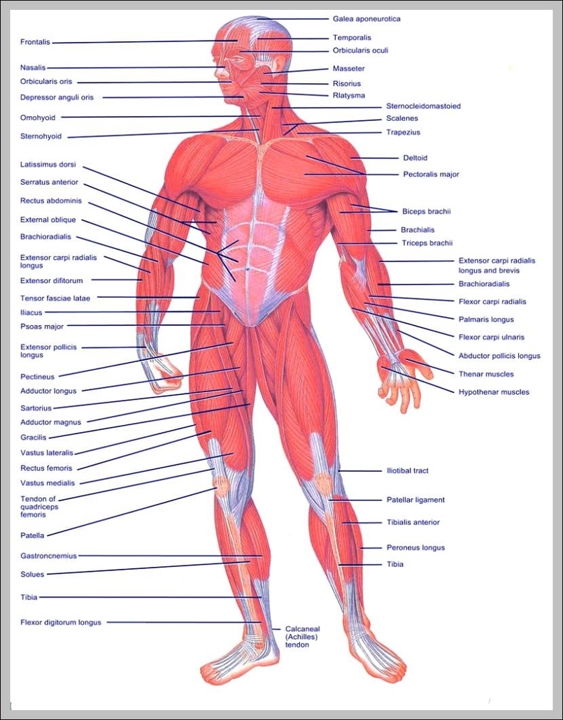
98 muscular system labeled diagram stock photos and images available, or start a new search to explore more stock photos and images. Frontal view of the muscular system of the male human body with descriptive labels pointing to the muscles on a white background. Labeled Anatomy Chart of Neck and Shoulder Muscles on White…

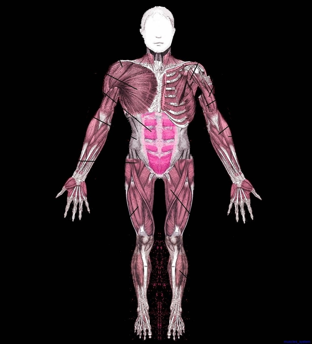
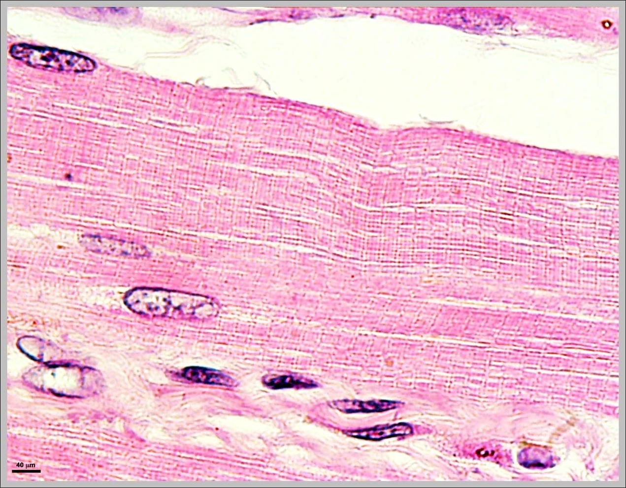
Last Updated: Jul 16, 2019 The muscular system is responsible for the movement of the human body. Attached to the bones of the skeletal system are about 700 named muscles that make up roughly half of a person’s body weight. Each of these muscles is a discrete organ constructed of skeletal muscle tissue, blood vessels, tendons, and nerves.

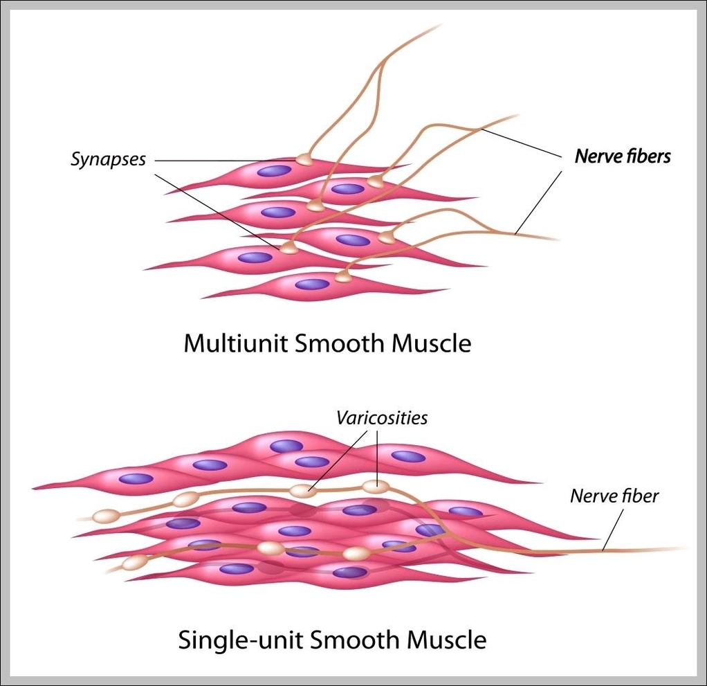
Human muscle system, the muscles of the human body that work the skeletal system, that are under voluntary control, and that are concerned with movement, posture, and balance. Broadly considered, human muscle—like the muscles of all vertebrates—is often divided into striated muscle (or skeletal muscle), smooth muscle, and cardiac muscle.

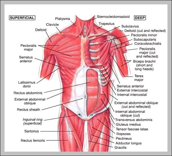
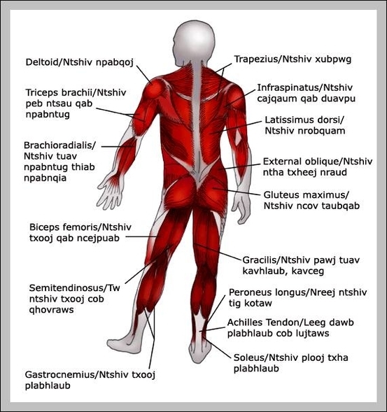
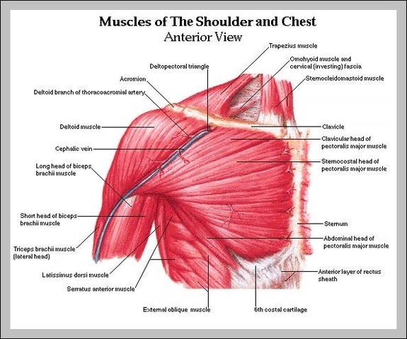
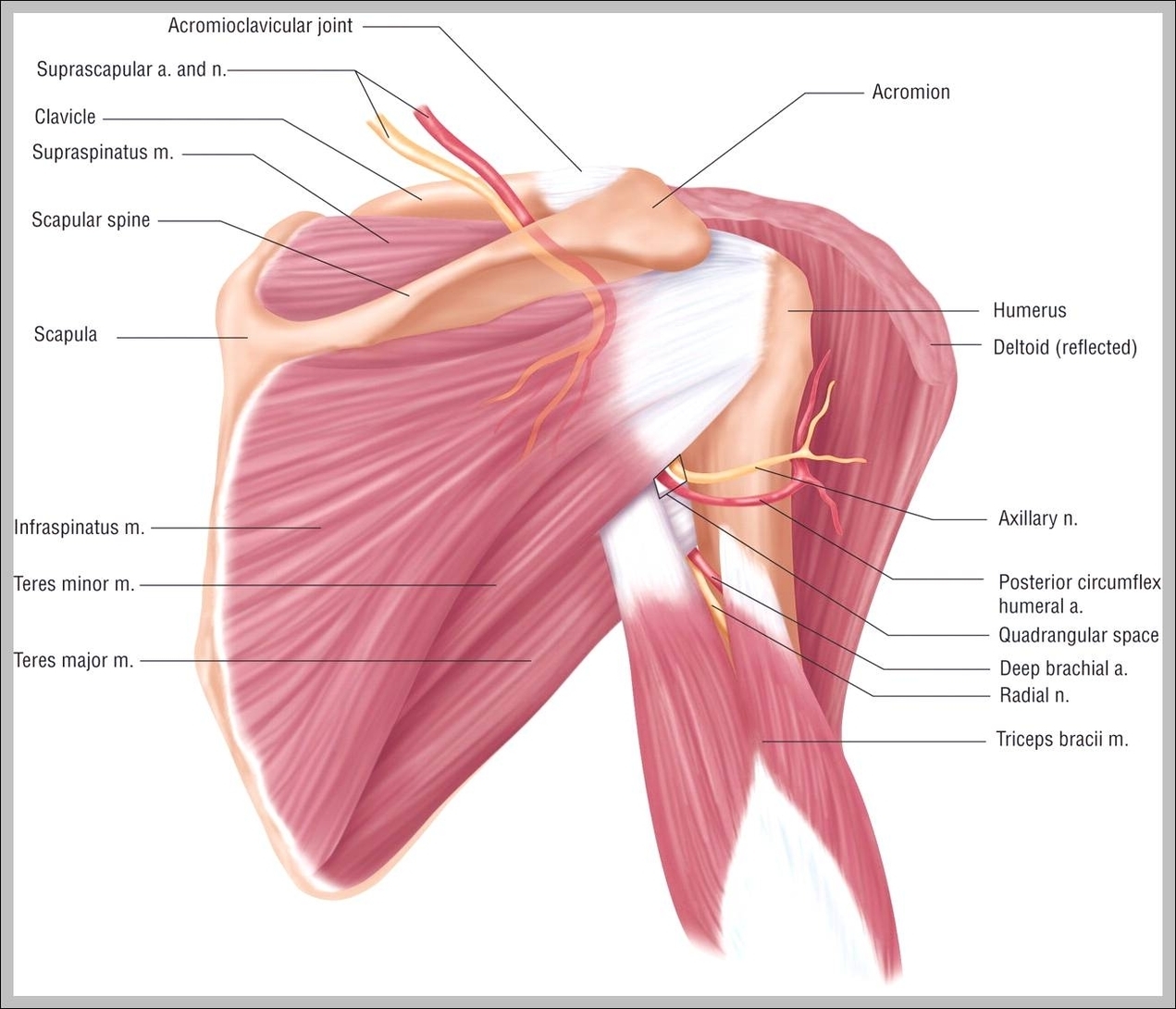
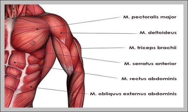
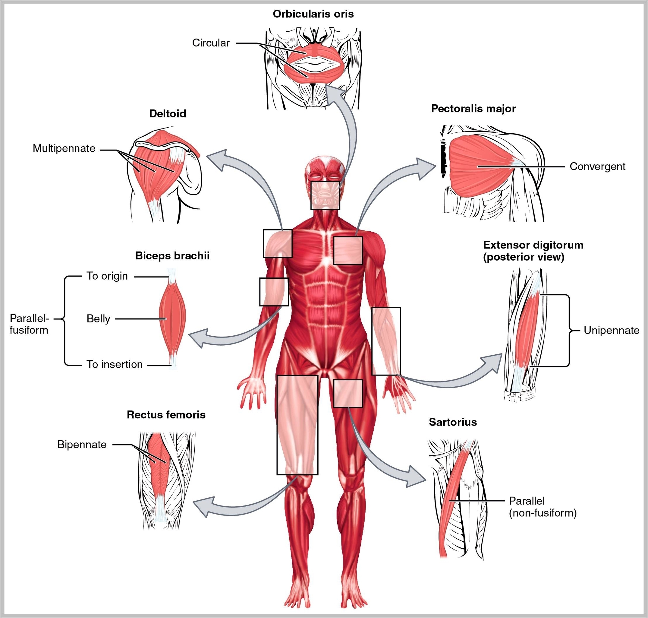
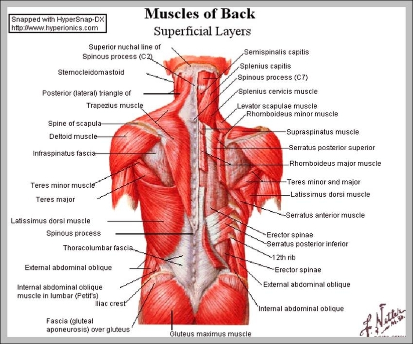
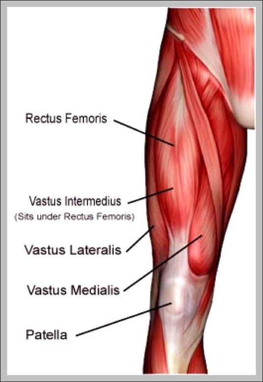
Muscles System Image Diagram - Chart - diagrams and charts with labels. This diagram depicts Muscles System Image

Muscles System Image 1

Nervous System Image

Nervous system Images and Stock Photos. 17,386 Nervous system photography and royalty free pictures available to download from thousands of stock photo providers. Human nervous system diagram. Pictures by Pixelchaos 52 / 978

Human nervous system medical vector illustration diagram with parasympathetic and sympathetic nerves and all connected inner organs through brain and spinal cord. Educational information complete guide. Woman suffering from bad headache or migraine. Appointment with doctor in office room. Sick and unwell lady with stress, trauma or burnout.

All of the bodies’ many sense organs are components of the nervous system. What are known as the special senses—vision, taste, smell, hearing, and balance—are all detected by specialized organs such as the eyes , taste buds , and olfactory epithelium.

Nervous System Image Diagram - Chart - diagrams and charts with labels. This diagram depicts Nervous System Image

Nervous System Image

Organs Of Skeletal System Image

What Are Human Skeletal System Organs? 1. Bones The most important organ of the skeletal system is the bones. Human skeleton is made up of 206 bones that in coordination not only provides support and protection to the viscera (with the help of muscles attached to them) but also produces blood cells for the body from the bone-marrow. 2.

19,988 human skeletal system stock photos, vectors, and illustrations are available royalty-free.

Regardless of age or sex, the skeletal system can be broken down into two parts, known as the axial skeleton and the appendicular skeleton. The adult axial skeleton consists of 80 bones. It’s made up of the bones that form the vertical axis of the body, such as the bones of the head, neck, chest, and spine.

Organs Of Skeletal System Image Diagram - Chart - diagrams and charts with labels. This diagram depicts Organs Of Skeletal System Image

Organs Of Skeletal System Image

Pancreatic System Image

Anatomy of the pancreas; drawing shows the pancreas, stomach, spleen, liver, gallbladder, bile ducts, colon, and small intestine. An inset shows the head, body, and tail of the pancreas. The bile duct and pancreatic duct are also shown. Anatomy of the pancreas. The pancreas has three areas: head, body, and tail.

1,908 pancreas anatomy stock photos and images available, or search for kidney anatomy or liver anatomy to find more great stock photos and pictures.

Pancreatitis: The pancreas becomes inflamed and damaged by its own digestive chemicals. Swelling and death of tissue of the pancreas can result. Although alcohol or gallstones can contribute, sometimes a cause for pancreatitis is never found. Pancreatic pseudocyst: After a bout of pancreatitis, a fluid-filled cavity called a pseudocyst can form.

Pancreatic System Image Diagram - Chart - diagrams and charts with labels. This diagram depicts Pancreatic System Image

Pancreatic System Image

Picture Of Female Reproductive System Diagram Image

227 female reproductive system diagram stock photos and images available, or start a new search to explore more stock photos and images. Teacher points to a diagram of female reproductive organs projected on a screen in a classroom in a scene from Human Growth, an education film on sex…

Female reproductive system. Genital organs. Human anatomy. Cervix, ovaries, fallopian tubes. Vector illustration. female reproductive system diagram stock illustrations Uterus. Female reproductive system. Genital organs. Human anatomy. Set of Female reproductive system uterus in different styles and cross sections. Front view in a cut.

3D computer graphic sagittal section of female reproductive system with labels. The female reproductive system (or female genital system) contains two main parts: the uterus, which hosts the developing fetus, produces vaginal and uterine secretions Anterior view of uterus, fallopian tube, ovary, cervix.

Picture Of Female Reproductive System Diagram Image Diagram - Chart - diagrams and charts with labels. This diagram depicts Picture Of Female Reproductive System Diagram Image

Picture Of Female Reproductive System Diagram Image scaled

Skeletal Muscles Diagram

Attached to the bones of the skeletal system are about 700 named muscles that make up roughly half of a person’s body weight. Each of these muscles is a discrete organ constructed of skeletal muscle tissue, blood vessels, tendons, and nerves.

This is a table of skeletal muscles of the human anatomy. There are around 640 skeletal muscles within the typical human body.

Cardiac and skeletal muscles are “striated” in that they contain sarcomeres and are packed into highly regular arrangements of bundles; smooth muscle has neither.

Skeletal Muscles Diagram Diagram - Chart - diagrams and charts with labels. This diagram depicts Skeletal Muscles Diagram

skeletal muscles diagram

Torso Muscles Diagram

The torso also harbours many of the main groups of muscles in the body, including the: 1 pectoral muscles 2 abdominal muscles 3 lateral muscle 4 epaxial muscles

The torso also harbours many of the main groups of muscles in the body, including the: The organs, muscles, and other contents of the torso are supplied by nerves, which mainly originate as nerve roots from the thoracic and lumbar parts of the spinal cord. Some organs also receive a nerve supply from the vagus nerve.

This is a table of skeletal muscles of the human anatomy. There are around 640 skeletal muscles within the typical human body.

Torso Muscles Diagram Diagram - Chart - diagrams and charts with labels. This diagram depicts Torso Muscles Diagram

torso muscles diagram

Pictures Of Muscle Cells

Types of muscle cells
Skeletal Muscle cells. Skeletal muscle cells are long, cylindrical, and striated.
Cardiac Muscle Cells. Cardiomyocytes are short and narrow, and fairly rectangular in shape.
Smooth Muscle Cells. Smooth muscle cells are spindle-shaped and contain a single central nucleus.

Muscle Cell Definition. A muscle cell, known technically as a myocyte, is a specialized animal cell which can shorten its length using a series of motor proteins specially arranged within the cell.

This structure is similar to the smooth endoplasmic reticulum of other types of cell. To produce contractile force, myosin associates with actin filaments, rotating a little and then pulling the filaments across each other, like oars propelling a boat. Skeletal muscle cells also contain two regulatory proteins, known as troponin and tropomyosin.

Pictures Of Muscle Cells Diagram - Chart - diagrams and charts with labels. This diagram depicts Pictures Of Muscle Cells

pictures of muscle cells

Skeletal Muscle Chart

skeletal muscle, which exists throughout the body The body’s collective muscle tissue constitutes its muscle mass. However, in most contexts, the term “muscle mass” specifically refers to skeletal muscle. This is the only type of muscle that a person can voluntarily control.

Skeletal muscle is a fascinating tissue with a complex structure. It consists of elongated multinuclear cells called the myocytes (or myofibers). The muscle cells can be anything from 1 mm to 30 cm in length. The longest muscle cell in our bodies can be found in the sartorius muscle and is 30 cm (nearly 12 inches!) long. From biology-forums.com

There are around 640 skeletal muscles within the typical human body. Almost every muscle constitutes one part of a pair of identical bilateral muscles, found on both sides, resulting in approximately 320 pairs of muscles, as presented in this article.

Skeletal Muscle Chart Diagram - Chart - diagrams and charts with labels. This diagram depicts Skeletal Muscle Chart

skeletal muscle chart

Picture Of Chest Muscles

The chest anatomy includes the pectoralis major, pectoralis minor and the serratus anterior. Learn about each of these muscles, their locations, functional anatomy and exercises for them. This page provides an overview of the chest muscle group.

Chest Anatomy: All About the Chest Muscles. The chest anatomy includes the pectoralis major, pectoralis minor and the serratus anterior. Learn about each of these muscles, their locations, functional anatomy and exercises for them.

Function of the Chest Muscles. The chest is part of a larger group of “pushing muscles” found in the upper body. The chest, as part of this group, enables you to perform pushing actions such as the barbell bench press or a daily activity such as moving a heavy dresser.

Picture Of Chest Muscles Diagram - Chart - diagrams and charts with labels. This diagram depicts Picture Of Chest Muscles

picture of chest muscles

Shoulder Muscle Chart 2

Toggle Anatomy System. On the anterior side of the shoulder, the coracobrachialis, serratus anterior, pectoralis major, and pectoralis minor muscles work as a group to flex and adduct the scapula and humerus anteriorly toward the sternum. The latissimus dorsi and teres major on the posterior side extend and adduct the arm towards the vertebrae…

Shoulder Muscles Anatomy. Shoulder muscles move the shoulder blades and upper arm bones. The main shoulder muscles are trapezius, deltoid, pectoralis major and 4 rotator cuff muscles: subscapularis, supraspinatus, infraspinatus and teres minor.

Each joint is surrounded by: tendons, to attach the muscles to the bones. The collection of muscles and tendons in the shoulder is known as the rotator cuff. It stabilizes the shoulder and holds the head of the humerus in the glenoid, a shallow cavity in the scapula.

Shoulder Muscle Chart 2 Diagram - Chart - diagrams and charts with labels. This diagram depicts Shoulder Muscle Chart 2

shoulder muscle chart 2

Pictures Of Shoulder Muscles

For that reason, and because of the dexterity of the shoulder joint itself, the musculature of the shoulder is complex, ranging from massive prime mover muscles to finer stabilizer and fixator muscles. The shoulder (or humeroscapular) joint is formed by the articulation of the head of the humerus with the scapula.

Medically reviewed by Healthline’s Medical Network on March 23, 2015. The shoulder has about eight muscles that attach to the scapula, humerus, and clavicle. These muscles form the outer shape of the shoulder and underarm.

The muscles in the shoulder aid in a wide range of movement and help protect and maintain the main shoulder joint, known as the glenohumeral joint. The largest of these shoulder muscles is the deltoid. This large triangular muscle covers the glenohumeral joint and gives the shoulder its rounded-off shape.

Pictures Of Shoulder Muscles Diagram - Chart - diagrams and charts with labels. This diagram depicts Pictures Of Shoulder Muscles

pictures of shoulder muscles

Upper Body Muscle Chart

Muscles. The major muscles in the upper torso of the body include: Trapezius: This muscle extends across the neck, shoulder, and back. It allows for movement of the shoulders and shoulder blades. Rhomboid major: Attached to the shoulder blade, this muscle is one of many that aids shoulder movement.

Muscle Anatomy: Human Anatomy Chart 1 Abs & Obliques Anatomy 2 Back Anatomy 3 Biceps Anatomy 4 Calves Anatomy 5 Chest Anatomy 6 Forearms Anatomy 7 Leg, Hip & Gluteal Anatomy 8 Neck Anatomy More …

Some compound exercises for the legs also involve the lower back. Bodybuilders commonly divide the body’s individual muscles into ten major muscle groups. These do not include the hip, neck and forearm muscles, which are rarely trained in isolation. The most common exercises for these muscle groups are listed above.

Upper Body Muscle Chart Diagram - Chart - diagrams and charts with labels. This diagram depicts Upper Body Muscle Chart

upper body muscle chart

Upper Body Muscle Names

List of Upper Body Muscles
Neck and Shoulders. The neck primarily consists of the sternocleidomastoid and the splenius muscles,…
Chest. The chest region includes two muscle groups and one individual muscle.
Abdominals. The abdominals primarily consist of the rectus abdominis, an individual muscle,…
Arms. The arms comprise two muscle groups…

The upper body comprises seven general muscle regions in the shoulders, chest and back. Each of these regions contains several muscle groups consisting of several muscle heads, or parts.

The main muscle group of the chest is the pectorals. A well-developed chest is very important because it will add major size to your inner upper body. The chest is a very visible part of the upper body, that can add weight and force to all your martial arts moves. The pectorals or pecs are the large chest muscles.

Upper Body Muscle Names Diagram - Chart - diagrams and charts with labels. This diagram depicts Upper Body Muscle Names

upper body muscle names

Upper Muscles Of The Body

Deltoids : The deltoids, also known as the shoulders contribute to both the front and back of the upper body. They are essential to building a strong upper body. Chest: The chest muscle is a major muscle in the upper body also. A well-developed chest is very important because it will add major size to your inner upper body.

Chest: The chest muscle is a major muscle in the upper body also. A well-developed chest is very important because it will add major size to your inner upper body.

The upper body comprises seven general muscle regions in the shoulders, chest and back. Each of these regions contains several muscle groups consisting of several muscle heads, or parts.

Upper Muscles Of The Body Diagram - Chart - diagrams and charts with labels. This diagram depicts Upper Muscles Of The Body

upper muscles of the body

Shoulder Muscles Pictures

For that reason, and because of the dexterity of the shoulder joint itself, the musculature of the shoulder is complex, ranging from massive prime mover muscles to finer stabilizer and fixator muscles. The shoulder (or humeroscapular) joint is formed by the articulation of the head of the humerus with the scapula.

Medically reviewed by Healthline’s Medical Network on March 23, 2015. The shoulder has about eight muscles that attach to the scapula, humerus, and clavicle. These muscles form the outer shape of the shoulder and underarm.

The muscles in the shoulder aid in a wide range of movement and help protect and maintain the main shoulder joint, known as the glenohumeral joint. The largest of these shoulder muscles is the deltoid. This large triangular muscle covers the glenohumeral joint and gives the shoulder its rounded-off shape.

Shoulder Muscles Pictures Diagram - Chart - diagrams and charts with labels. This diagram depicts Shoulder Muscles Pictures

shoulder muscles pictures

Skeletal Muscle Picture 2

Skeletal muscle, also called voluntary muscle, in vertebrates, most common of the three types of muscle in the body. Skeletal muscles are attached to bones by tendons, and they produce all the movements of body parts in relation to each other. Unlike smooth muscle and cardiac muscle, skeletal muscle is under voluntary control.

Skeletal muscle, also called voluntary muscle, in vertebrates, most common of the three types of muscle in the body.

[edit on Wikidata] Skeletal muscle is one of three major muscle types, the others being cardiac muscle and smooth muscle. It is a form of striated muscle tissue, which is under the voluntary control of the somatic nervous system. Most skeletal muscles are attached to bones by bundles of collagen fibers known as tendons.

Skeletal Muscle Picture 2 Diagram - Chart - diagrams and charts with labels. This diagram depicts Skeletal Muscle Picture 2

skeletal muscle picture 2

Types Of Muscle Cells

Types of muscle cells
Skeletal Muscle cells. Skeletal muscle cells are long, cylindrical, and striated.
Cardiac Muscle Cells. Cardiomyocytes are short and narrow, and fairly rectangular in shape.
Smooth Muscle Cells. Smooth muscle cells are spindle-shaped and contain a single central nucleus.

The 3 types of muscle tissue are cardiac, smooth, and skeletal. Cardiac muscle cells are located in the walls of the heart, appear striated, and are under involuntary control.

Skeletal muscle cells 1 Muscle cells, commonly known as myocytes, are the cells that make up muscle tissue. There are 3 types of muscle cells in… 2 Skeletal muscle cells are long, cylindrical, multi-nucleated and striated . Each nucleus regulates the metabolic… More …

Types Of Muscle Cells Diagram - Chart - diagrams and charts with labels. This diagram depicts Types Of Muscle Cells

types of muscle cells

Rectus Muscle

Rectus abdominis. The human rectus abdominis muscle. [edit on Wikidata] Example of a man with a visible rectus abdominis. The rectus abdominis muscle, also known as the “abdominal muscle” or “abs”, is a paired muscle running vertically on each side of the anterior wall of the human abdomen, as well as that of some other mammals.

Rectus abdominis. The rectus abdominis muscle is located in the front of the body, beginning at the pubic bone and ending at the sternum. It is located inside the abdominal region. The muscle is activated while doing crunches because it pulls the ribs and the pelvis in and curves the back.

The rectus abdominis is an important postural muscle. It is responsible for flexing the lumbar spine, as when doing a so-called “crunch” sit up.

Rectus Muscle Diagram - Chart - diagrams and charts with labels. This diagram depicts Rectus Muscle

rectus muscle