Pictures Of Bones In The Human Body

List of bones of the human skeleton. The human skeleton of an adult consists of 206-208 bones. It is composed of 270 bones at birth, but later decreases to 80 bones in the axial skeleton and 126 bones in the appendicular skeleton. Many small supernumerary bones, such as some sesamoid bones, are not included in this count.

List of Bones in the Human Body. 1 Frontal Bone. This bone forms the forehead, the roof of the orbital cavity (eye socket), and the root of the nose. A newborn has a frontal bone that … 2 Parietal Bones. 3 Temporal Bones. 4 Occipital Bone. 5 Sphenoid Bone. More items

However, as a child grows, some of the bones fuse together. The result is that there are 206 bones in the body of an adult human being. This difference in the number of bones helps forensic anthropologists in determining the age of an individual through the skeletal remains, mainly the skull.

Pictures Of Bones In The Human Body Diagram - Chart - diagrams and charts with labels. This diagram depicts Pictures Of Bones In The Human Body

pictures of bones in the human body

Upper Muscles Of The Body

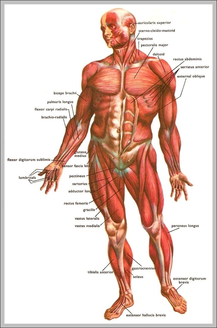
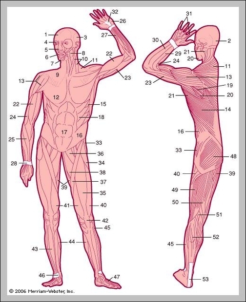
Deltoids : The deltoids, also known as the shoulders contribute to both the front and back of the upper body. They are essential to building a strong upper body. Chest: The chest muscle is a major muscle in the upper body also. A well-developed chest is very important because it will add major size to your inner upper body.

Chest: The chest muscle is a major muscle in the upper body also. A well-developed chest is very important because it will add major size to your inner upper body.

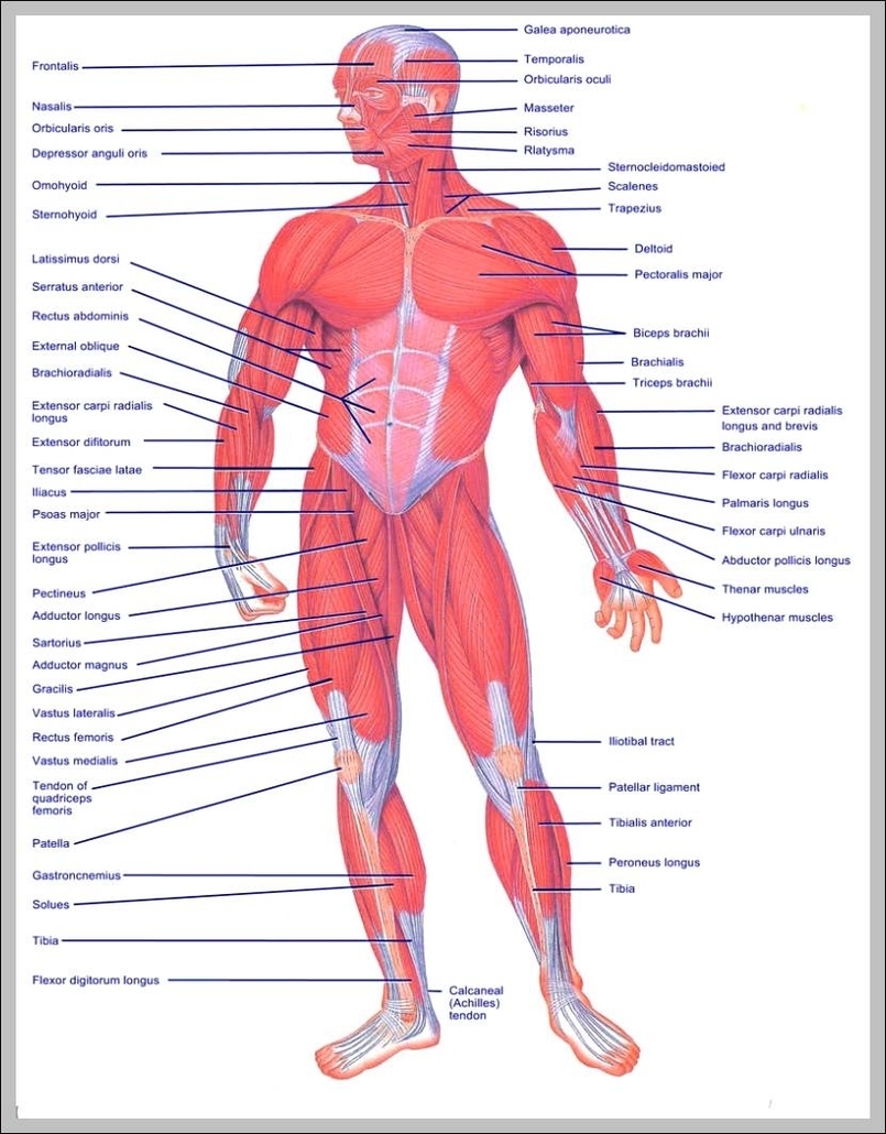
The upper body comprises seven general muscle regions in the shoulders, chest and back. Each of these regions contains several muscle groups consisting of several muscle heads, or parts.

Upper Muscles Of The Body Diagram - Chart - diagrams and charts with labels. This diagram depicts Upper Muscles Of The Body

upper muscles of the body

Where Is The Kidney Located In The Human Body

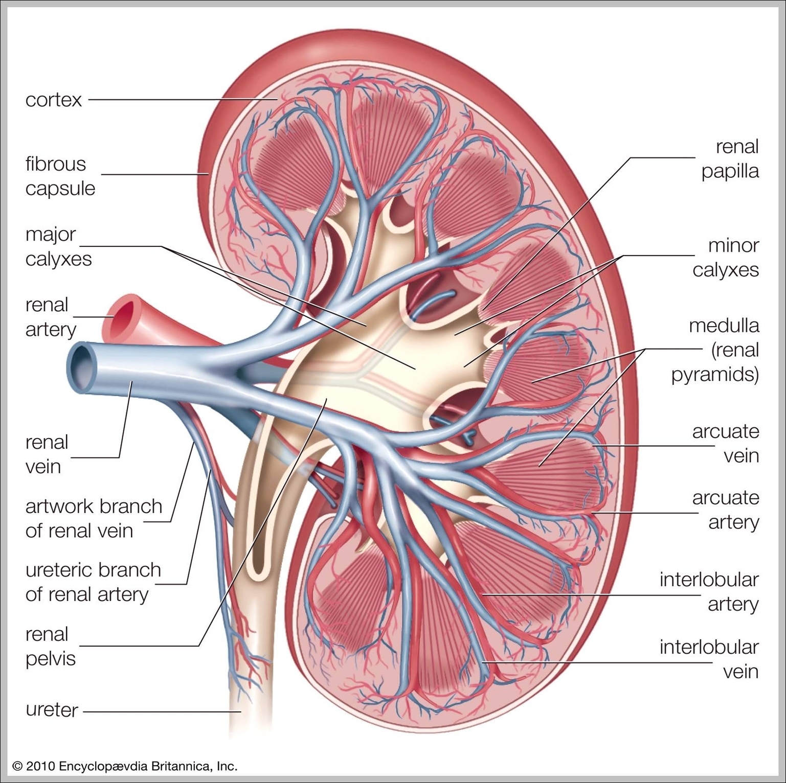
The kidneys are a pair of bean-shaped organs on either side of your spine, below your ribs and behind your belly. Each kidney is about 4 or 5 inches long …

The kidneys are recognized as the major organs of the urinary system. The kidneys are two small organs that are located near the vertebral column at the posterior or the back portion of the body. Both kidneys are located on opposite sides; however, the one of the left is placed a little higher compared to the one on the right.

The kidneys are bean-shaped and are located high in the abdominal cavity, one on each side of the spine, and lie in a retroperitoneal position at a slightly oblique angle. The right kidney sits just below the diaphragm and posterior to the liver.

Where Is The Kidney Located In The Human Body Diagram - Chart - diagrams and charts with labels. This diagram depicts Where Is The Kidney Located In The Human Body

where is the kidney located in the human body

Pictures Of Body Parts

Human Body Parts List. Picture of Internal Organs. Parts of the Body. 1. head 2. arm 3. back 4. waist 5. buttocks/ backside 6. leg 7. face 8. chest 9. stomach 10. hip 11. hand 12. foot. 13. eye 14. eyebrow 15. nose 16. mouth 17. chin 18. hair 19. ear 20. lips 21. neck. 22. nail 23. thumb 24. finger 25. wrist

Parts of the Body Girl. eye, nose, cheek, chin, mouth, neck, shoulder, armpit, breast, thorax, navel, abdomen, publs, groin, knee, foot, ankle, toe. HUMAN BODY WOMAN POSTERIOR VIEW. hair. nape, head, neck, shoulder blade, arm, elbow, back, waist, trunk, loin, hip, forearm, wrist, hand, buttock, thigh, leg, calf, foot, heel.

Picture of Internal Organs. Parts of the Body. 1. head 2. arm 3. back 4. waist 5. buttocks/ backside 6. leg 7. face 8. chest 9. stomach 10. hip 11. hand 12. foot. 13. eye 14. eyebrow 15. nose 16. mouth 17. chin 18. hair 19. ear 20. lips 21. neck. 22. nail 23. thumb 24. finger 25. wrist

Pictures Of Body Parts Diagram - Chart - diagrams and charts with labels. This diagram depicts Pictures Of Body Parts

pictures of body parts

Picture Of a Human Body

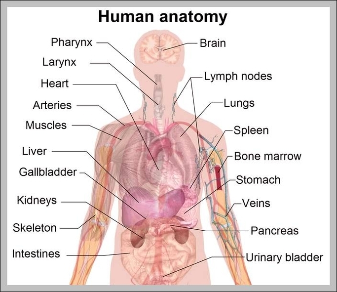
Photo description: This diagram of the human body shows a range of organs that are important to human anatomy. They include the brain, heart, lungs, spleen, muscles, stomach, kidneys and more.

Parts of the Body, Human body parts: Name and Pictures 1 HEAD PARTS 2 FACE PART NAMES 3 HAND PARTS NAME 4 FOOT PARTS NAMES 5 Body Parts Pictures for Classroom and Therapy More …

Parts of the Body Girl eye, nose, cheek, chin, mouth, neck, shoulder, armpit, breast, thorax, navel, abdomen, publs, groin, knee, foot, ankle, toe HUMAN BODY WOMAN POSTERIOR VIEW hair. nape, head, neck, shoulder blade, arm, elbow, back, waist, trunk, loin, hip, forearm, wrist, hand, buttock, thigh, leg, calf, foot, heel.

Picture Of a Human Body Diagram - Chart - diagrams and charts with labels. This diagram depicts Picture Of a Human Body

picture of a human body

Where Is The Liver Located In The Body

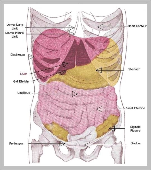
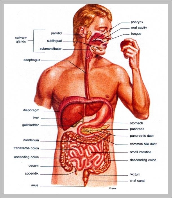
Liver Location. The liver is located in the upper right quadrant of the abdominal cavity, right below the diaphragm. It sits above the right kidney, intestine and the stomach. The rib cage protects this organ. The esophagus or the food pipe passes posteriorly of the organ and enters the stomach situated below it.

Your liver is located near your abdomen on the right side, actually beneath your ribs and above your diaphragm which separates the chest and the abdomen. The liver is a gland but it is also the largest solid organ in the body.

Where Is Your Liver Located And What Causes Liver Pain. Your liver is located on the right side of your body in the upper right abdomen below your ribcage. The liver is one of the largest internal organs in your body and its main function is to filter blood and remove toxins.

Where Is The Liver Located In The Body Diagram - Chart - diagrams and charts with labels. This diagram depicts Where Is The Liver Located In The Body

where is the liver located in the body

Where Is The Heart In The Human Body

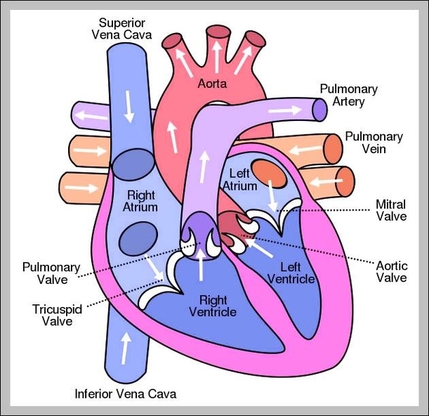
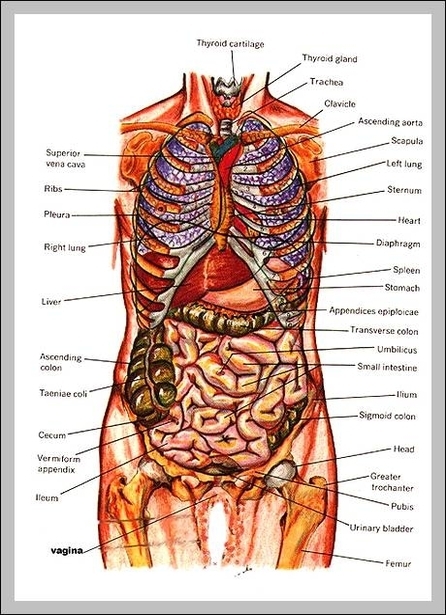
The heart is a muscular organ about the size of a fist, located just behind and slightly left of the breastbone. The heart pumps blood through the network of arteries and veins called the cardiovascular system.

In general, science tells us that the heart situated very close to the center of our chest. One-third part of our heart is on the right side, while two-third located on the left side of our chest. It means it’s pretty simple to say that it’s in the center of our chest.

The inferior tip of the heart, known as the apex, rests just superior to the diaphragm. The base of the heart is located along the body’s midline with the apex pointing toward the left side. Because the heart points to the left, about 2/3 of the heart’s mass is found on the left side of the body and the other 1/3 is on the right.

Where Is The Heart In The Human Body Diagram - Chart - diagrams and charts with labels. This diagram depicts Where Is The Heart In The Human Body

where is the heart in the human body

Where Are Organs In Your Body

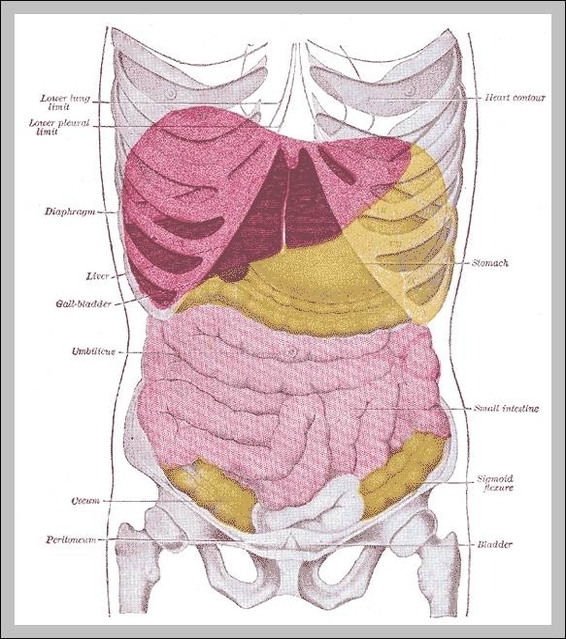
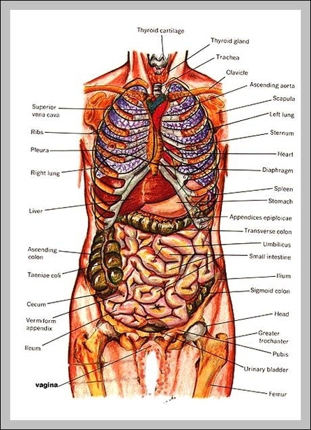
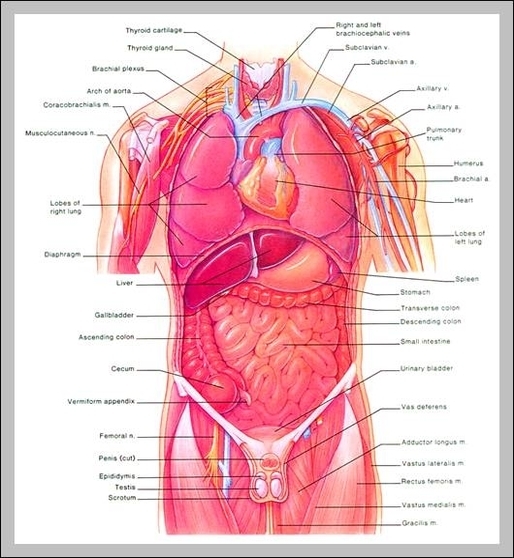
While a few organs are located in the near central plane, the position of most organs is either to the left or right side of the body.

Your body organs range from your brain, heart, liver, skin, lungs, kidneys, intestines, stomach, bladder, etc. In all, there are believed to be 80 organs in your body all serving different functions and uses.

It is located on the right side of the body above the stomach, as seen in the image above. It is an essential organ of metabolism and one of the principal organs in the human body that detoxifies substances. It helps to convert any toxic substances or drugs into water-soluble inert materials. These are then readily excreted by the kidneys.

Where Are Organs In Your Body Diagram - Chart - diagrams and charts with labels. This diagram depicts Where Are Organs In Your Body

where are organs in your body

Show Picture Of Human Body

Photo description: This diagram of the human body shows a range of organs that are important to human anatomy. They include the brain, heart, lungs, spleen, muscles, stomach, kidneys and more.

Parts of the Body, Human body parts: Name and Pictures 1 HEAD PARTS 2 FACE PART NAMES 3 HAND PARTS NAME 4 FOOT PARTS NAMES 5 Body Parts Pictures for Classroom and Therapy More …

These four basic cell types, together with their extracellular materials, form the fundamental tissues of the human body: (1) epithelial tissues, which cover the body’s surface and line the internal organs, body cavities, and passageways; (2) muscle tissues]

Show Picture Of Human Body Diagram - Chart - diagrams and charts with labels. This diagram depicts Show Picture Of Human Body

show picture of human body

Picture Of Inside Body

The brain regulates breathing, body temperature and allows you to think, speak, imagine and experience emotions. After the brain, the lungs are the fourth largest organ. Here a smoker’s dissected heart (center) with the lungs surrounding it, showing black spots from tar.

Parts of the Body Girl eye, nose, cheek, chin, mouth, neck, shoulder, armpit, breast, thorax, navel, abdomen, publs, groin, knee, foot, ankle, toe HUMAN BODY WOMAN POSTERIOR VIEW hair. nape, head, neck, shoulder blade, arm, elbow, back, waist, trunk, loin, hip, forearm, wrist, hand, buttock, thigh, leg, calf, foot, heel.

Internal organs of the human body vocabulary. Brain. uvula. Pharynx. Spinal cord. Tonsil. Gullet / esophagus. larynx. Windpipe / Trachea. Bronchial tube. Heart.

Picture Of Inside Body Diagram - Chart - diagrams and charts with labels. This diagram depicts Picture Of Inside Body

picture of inside body

Pineal Body

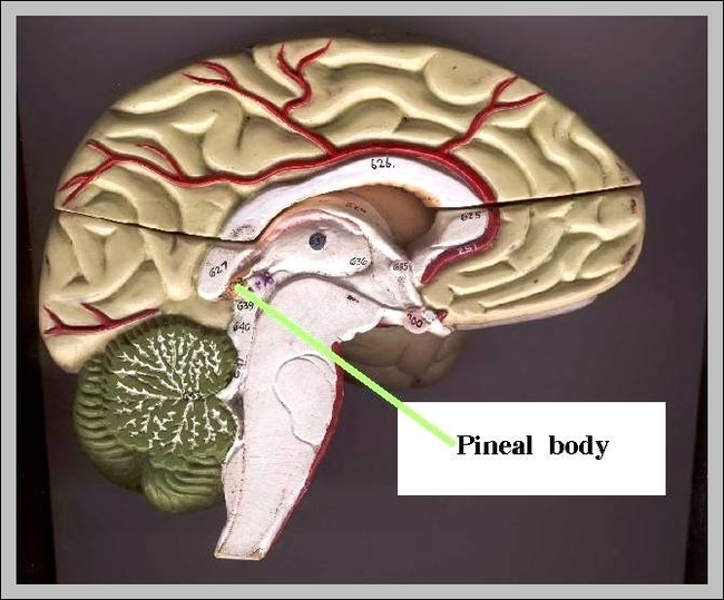
Basic Pineal Body Function. The pineal gland is one of the most important endocrine glands. Further, it regulates other glands as well, such as pituitary and thyroid. The essential function of the pineal gland is to secrete melatonin. And that, in turn, impacts the central nervous systems greatly.

Unlike most of the mammalian brain, the pineal gland is not isolated from the body by the blood–brain barrier system; it has profuse blood flow, second only to the kidney, supplied from the choroidal branches of the posterior cerebral artery.

The pineal gland is a small endocrine gland in the brain of animals with backbones. The pineal gland produces melatonin, a serotonin-derived hormone which modulates sleep patterns in both circadian and seasonal cycles. The shape of the gland resembles a pine cone from which it derived its name.

Pineal Body Diagram - Chart - diagrams and charts with labels. This diagram depicts Pineal Body

pineal body

Picture Of Internal Organs Of The Human Body

Human body internal organs. Stomach and lungs, kidneys and heart, brain and liver. Vector isolated illustration of human internal organs in female body. Stomach, liver, intestine, bladder, lung, testicle, uterus, spine, pancreas, kidney, heart, bladder icon.

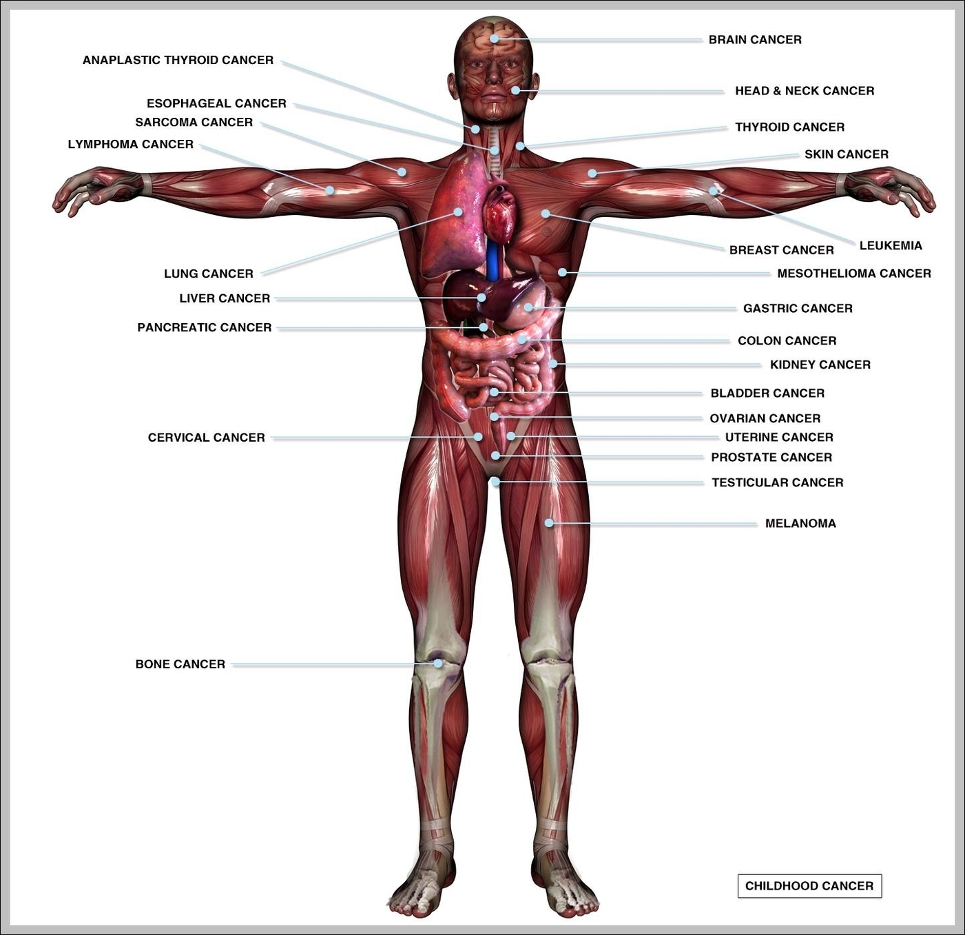
human internal organ images 101,663 human internal organ stock photos, vectors, and illustrations are available royalty-free. See human internal organ stock video clips of 1,017 internal organsbrain heartintestine symboljoints musclesanatomy organshead and neck cancersbody organsuterus linebrain heart boneinternal organ

Your body organs range from your brain, heart, liver, skin, lungs, kidneys, intestines, stomach, bladder, etc.

Picture Of Internal Organs Of The Human Body Diagram - Chart - diagrams and charts with labels. This diagram depicts Picture Of Internal Organs Of The Human Body

picture of internal organs of the human body

Tendons In The Body

Tendon, tissue that attaches a muscle to other body parts, usually bones. Tendons are the connective tissues that transmit the mechanical force of muscle contraction to the bones; the tendon is firmly connected to muscle fibres at one end and to components of the bone at its other end. Tendons are

Tendons are made out collagen that supplies them with strength and nutrition. Tendons work along with the muscles, and they are found all over the body. It has to stay with the muscle to protect and keep it connected to the bone. There are several important tendons in the body, and some of them are discussed below.

Tendons In The Body Diagram - Chart - diagrams and charts with labels. This diagram depicts Tendons In The Body

tendons in the body

Pictures Inside The Human Body

Parts of the Body, Human body parts: Name and Pictures 1 HEAD PARTS 2 FACE PART NAMES 3 HAND PARTS NAME 4 FOOT PARTS NAMES 5 Body Parts Pictures for Classroom and Therapy More …

The brain regulates breathing, body temperature and allows you to think, speak, imagine and experience emotions. After the brain, the lungs are the fourth largest organ. Here a smoker’s dissected heart (center) with the lungs surrounding it, showing black spots from tar.

Parts of the Body Girl eye, nose, cheek, chin, mouth, neck, shoulder, armpit, breast, thorax, navel, abdomen, publs, groin, knee, foot, ankle, toe HUMAN BODY WOMAN POSTERIOR VIEW hair. nape, head, neck, shoulder blade, arm, elbow, back, waist, trunk, loin, hip, forearm, wrist, hand, buttock, thigh, leg, calf, foot, heel.

Pictures Inside The Human Body Diagram - Chart - diagrams and charts with labels. This diagram depicts Pictures Inside The Human Body

pictures inside the human body

Vein System Of The Human Body

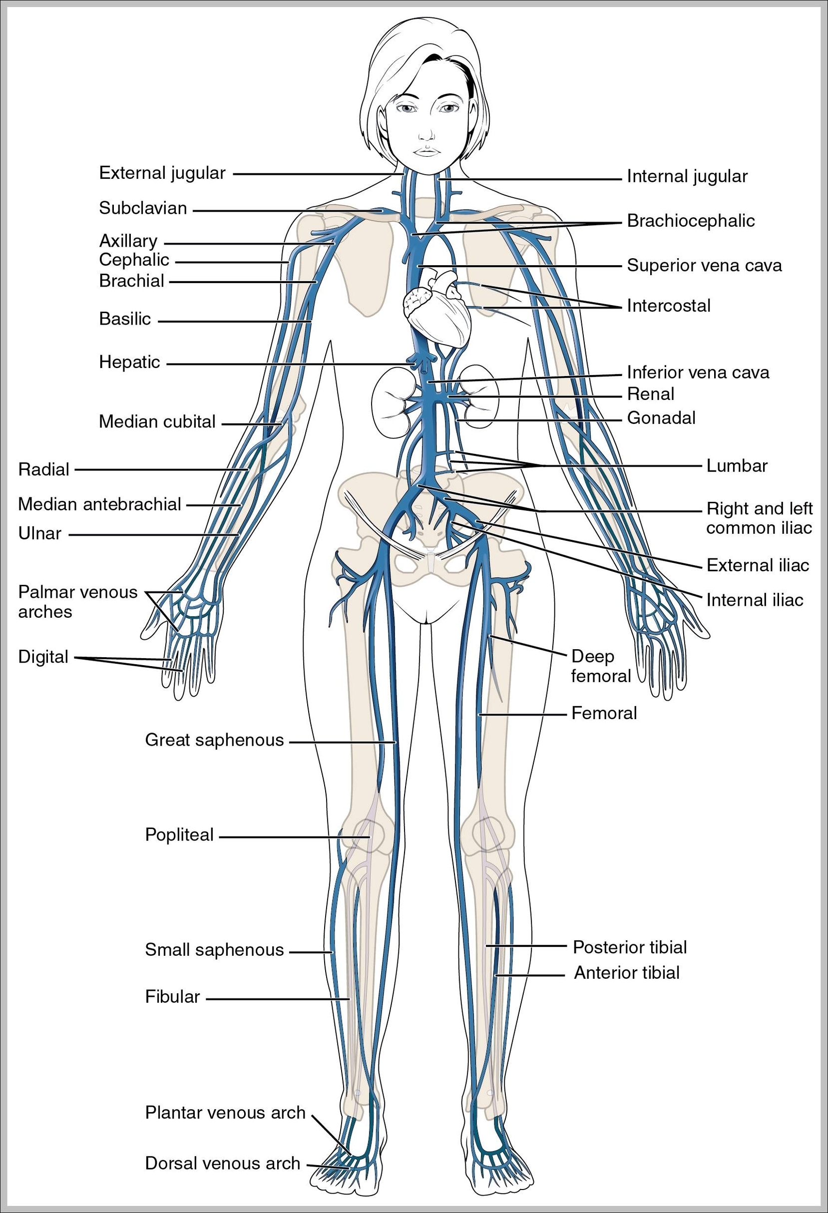
The largest veins in the human body are the venae cavae. These are two large veins which enter the right atrium of the heart from above and below. The superior vena cava carries blood from the arms and head to the right atrium of the heart, while the inferior vena cava carries blood from the legs and abdomen to the heart.

The venules branch into larger veins which eventually carry the blood to the largest veins in the body, the vena cava. Blood is then transported from the superior vena cava and inferior vena cava to the right atrium of the heart. Veins are composed of layers of thin tissue. The vein wall consists of three layers:

It’s a single layer of endothelium cells and some connective tissue. This layer sometimes contains one-way valves, especially in the veins of your arms and legs. These valves prevent blood from flowing backward. Veins are often categorized based on their location and any unique features or functions.

Vein System Of The Human Body Diagram - Chart - diagrams and charts with labels. This diagram depicts Vein System Of The Human Body

vein system of the human body

Veins In The Body Diagram

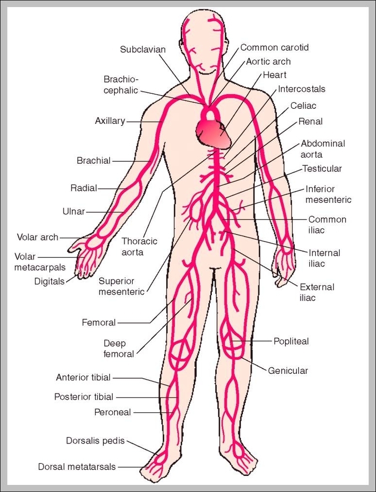
Major arteries, veins and nerves of the body. 1 Head and neck. Veins and arteries of the head (a diagram) 2 Trunk. Veins of the trunk converge from the thorax, abdomen, and pelvis towards the heart. Deoxygenated blood from the thorax ultimately drains into … 3 Upper extremity. 4 Lower extremity.

It’s a single layer of endothelium cells and some connective tissue. This layer sometimes contains one-way valves, especially in the veins of your arms and legs. These valves prevent blood from flowing backward. Veins are often categorized based on their location and any unique features or functions.

Blood from superficial veins is often directed into the deep veins through short veins called connecting veins. Valves in these veins allow blood to flow from the superficial veins to your deep veins, but not the other way. Use this interactive 3-D diagram to explore the venous system.

Veins In The Body Diagram Diagram - Chart - diagrams and charts with labels. This diagram depicts Veins In The Body Diagram

veins in the body diagram