Anatomy Brain Nervous Ffbb Efecbdlarge Image

WebMD’s Brain Anatomy Page provides a detailed diagram and definition of the brain including its function, parts, and conditions that affect it. Skip to main content Check Your Symptoms Find A Doctor Find A Dentist Connect to Care Find Lowest Drug Prices Health A-Z Health A-Z Common Conditions ADD/ADHD Allergies

The cerebral cortex is what we see when we look at the brain. It is the outermost portion that can be divided into the four lobes of the brain. Each bump on the surface of the brain is known as a gyrus, while each groove is known as a sulcus.

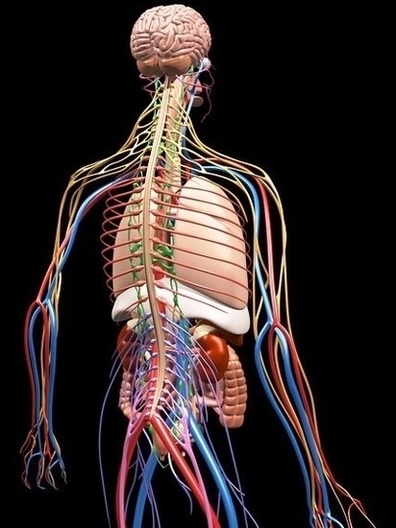
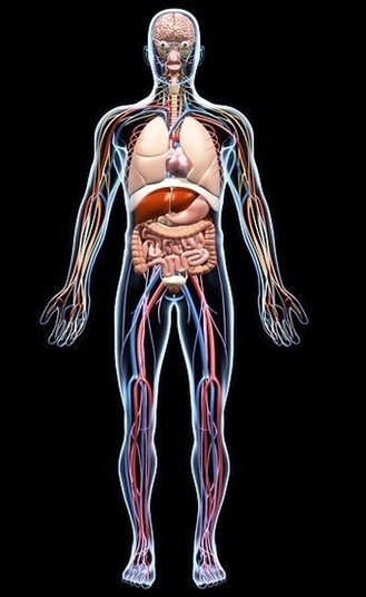
The brain is made up of billions of neurons and that it also has a number of specialized parts that are each involved in important functions. While there is still a great deal that researchers do not yet know about the brain, they have learned a great deal about the anatomy and function of the brain.

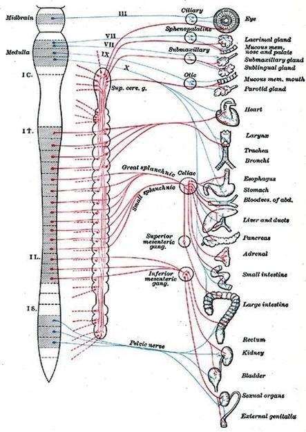
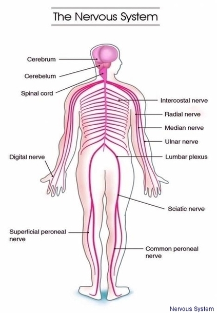
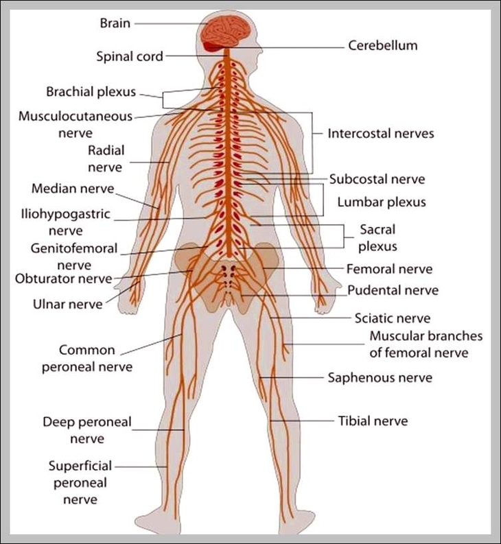
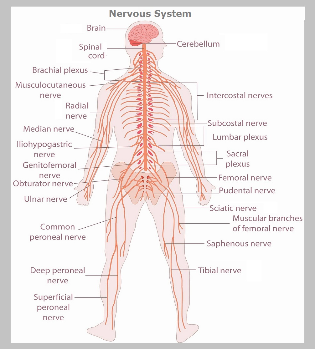
Anatomy Brain Nervous Ffbb Efecbdlarge Image Diagram - Chart - diagrams and charts with labels. This diagram depicts Anatomy Brain Nervous Ffbb Efecbdlarge Image

Anatomy Brain Nervous Ffbb Efecbdlarge Image

Autonomic Nervous System Image

Autonomic nervous system (ANS) is a functional division of the nervous system, with its structural parts in both the central nervous system (CNS) and the peripheral nervous system (PNS). It controls the glands and smooth muscle of all the internal organs (viscera) unconsciously.

The hypothalamus, just above the brain stem, acts as an integrator for autonomic functions, receiving ANS regulatory input from the limbic system to do so. The autonomic nervous system has three branches: the sympathetic nervous system, the parasympathetic nervous system and the enteric nervous system.

The autonomic nervous system is unique in that it requires a sequential two-neuron efferent pathway; the preganglionic neuron must first synapse onto a postganglionic neuron before innervating the target organ.

Autonomic Nervous System Image Diagram - Chart - diagrams and charts with labels. This diagram depicts Autonomic Nervous System Image

Autonomic Nervous System Image

Peripheral Nervous System Image

The peripheral nervous system (PNS)is the division of the nervous systemcontaining all the nerves that lie outside of the central nervous system (CNS). The primary role of the PNS is to connect the CNS to the limbs, and skin.

Our nervous system is divided in two components: the central nervous system(CNS), which includes the brain and spinal cord, and the peripheral nervous system (PNS), which encompasses nerves outside the brain and spinal cord. These two components cooperate at all times to ensure our lively functions: we are nothing without our nervous system!

The primary role of the PNS is to connect the CNS to the limbs, and skin. These nerves extend from the central nervous system to the outermost areas of the body. The peripheral system allows the brain and spinal cord to receive and send information to other areas of the body, which allows us to react to stimuli in our environment.

Peripheral Nervous System Image Diagram - Chart - diagrams and charts with labels. This diagram depicts Peripheral Nervous System Image

Peripheral Nervous System Image

Anatomy Brain Nervous Fee Db Fa Ecffffclarge Image

Anatomy of the Brain The anatomy of the brain is complex due its intricate structure and function. This amazing organ acts as a control center by receiving, interpreting, and directing sensory information throughout the body. The brain and spinal cord are the two main structures of the central nervous system.

The anatomy of the brain is complex due its intricate structure and function. This amazing organ acts as a control center by receiving, interpreting, and directing sensory information throughout the body.

Limbic System Structures. Amygdala – involved in emotional responses, hormonal secretions, and memory. Cingulate Gyrus – a fold in the brain involved with sensory input concerning emotions and the regulation of aggressive behavior.

Anatomy Brain Nervous Fee Db Fa Ecffffclarge Image Diagram - Chart - diagrams and charts with labels. This diagram depicts Anatomy Brain Nervous Fee Db Fa Ecffffclarge Image

Anatomy Brain Nervous Fee Db Fa Ecffffclarge Image

Nervous Main Image

Last Updated: Jun 15, 2018. The nervous system consists of the brain, spinal cord, sensory and all of the nerves that connect these organs with the rest of the body. Together, these organs are responsible for the control of the body and communication among its parts.

Functions of the Nervous System. The nervous system has 3 main functions: sensory, integration, and motor. Sensory. The sensory function of the nervous system involves collecting information from sensory receptors that monitor the body’s internal and external conditions.

1 Afferent, Efferent, and Mixed Nerves. Some of the nerves in the body are specialized for carrying information in only one direction, similar to a one-way street. … 2 Cranial Nerves. Extending from the inferior side of the brain are 12 pairs of cranial nerves. … 3 Spinal Nerves. …

Nervous Main Image Diagram - Chart - diagrams and charts with labels. This diagram depicts Nervous Main Image

Nervous Main Image

Nervous System Image

Nervous system Images and Stock Photos. 17,386 Nervous system photography and royalty free pictures available to download from thousands of stock photo providers. Human nervous system diagram. Pictures by Pixelchaos 52 / 978

Human nervous system medical vector illustration diagram with parasympathetic and sympathetic nerves and all connected inner organs through brain and spinal cord. Educational information complete guide. Woman suffering from bad headache or migraine. Appointment with doctor in office room. Sick and unwell lady with stress, trauma or burnout.

All of the bodies’ many sense organs are components of the nervous system. What are known as the special senses—vision, taste, smell, hearing, and balance—are all detected by specialized organs such as the eyes , taste buds , and olfactory epithelium.

Nervous System Image Diagram - Chart - diagrams and charts with labels. This diagram depicts Nervous System Image

Nervous System Image

Nervous System

Nervous System – Nervous System Diagram – Nervous System Chart – Human nervous system anatomy consists of the following parts brain, nerves, spinal cord, lumbar plexus, ulnar nerve and other types of nerves. Human nervous system diagram with labels.

nervous system

Nervous System Diagram - Chart - diagrams and charts with labels. This diagram depicts Nervous System

nervous system diagram