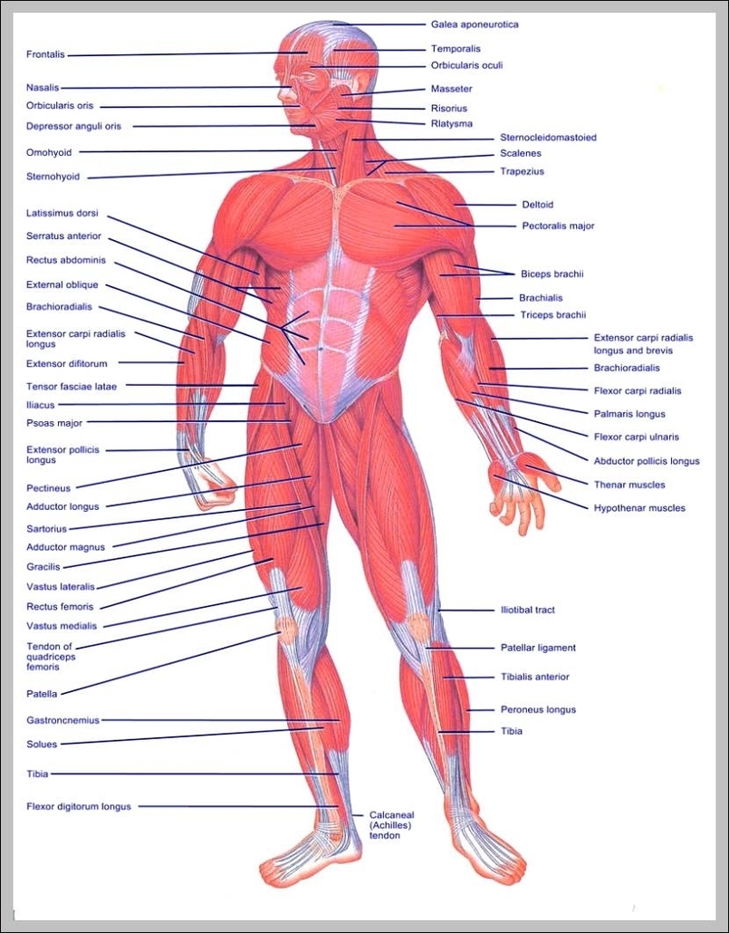
List of Upper Body Muscles
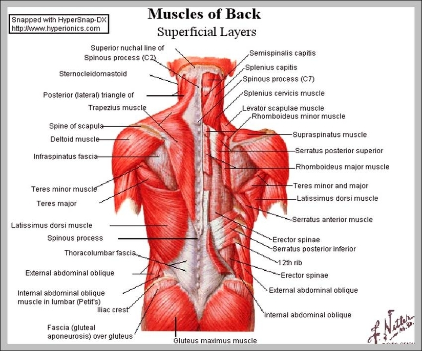
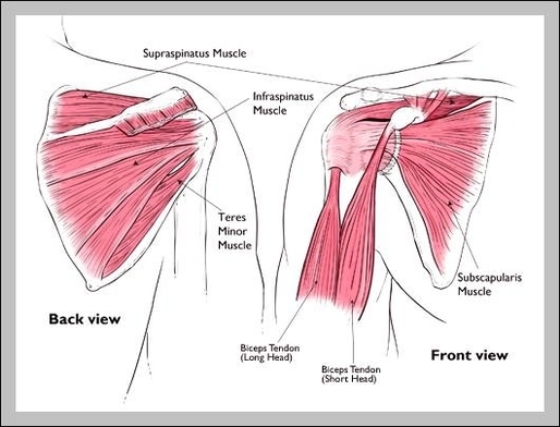
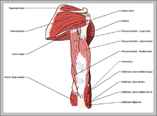
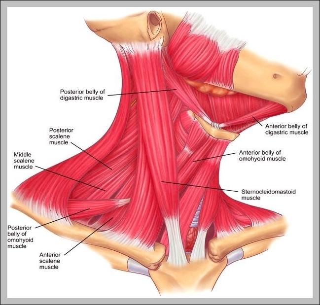
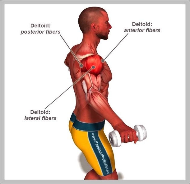
Neck and Shoulders. The neck primarily consists of the sternocleidomastoid and the splenius muscles,…
Chest. The chest region includes two muscle groups and one individual muscle.
Abdominals. The abdominals primarily consist of the rectus abdominis, an individual muscle,…
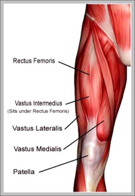
Arms. The arms comprise two muscle groups…
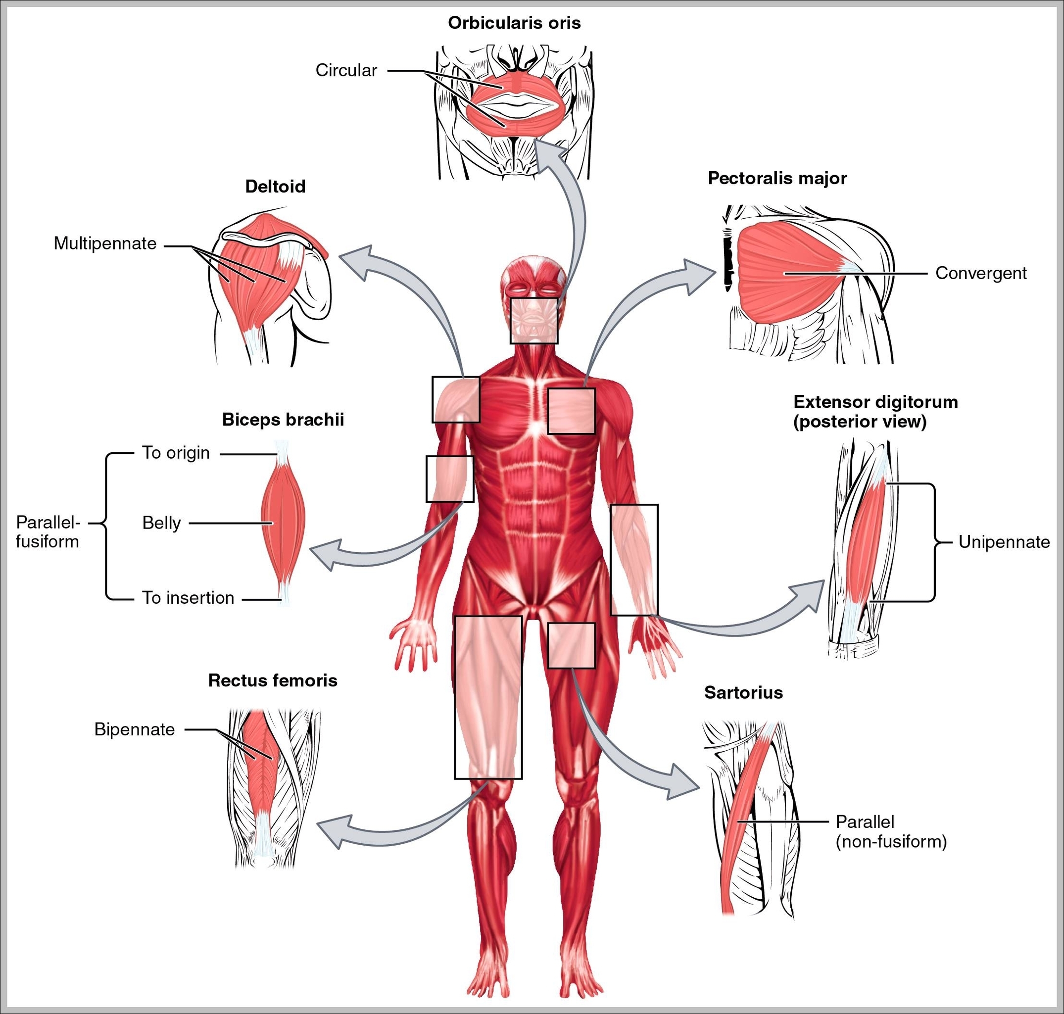
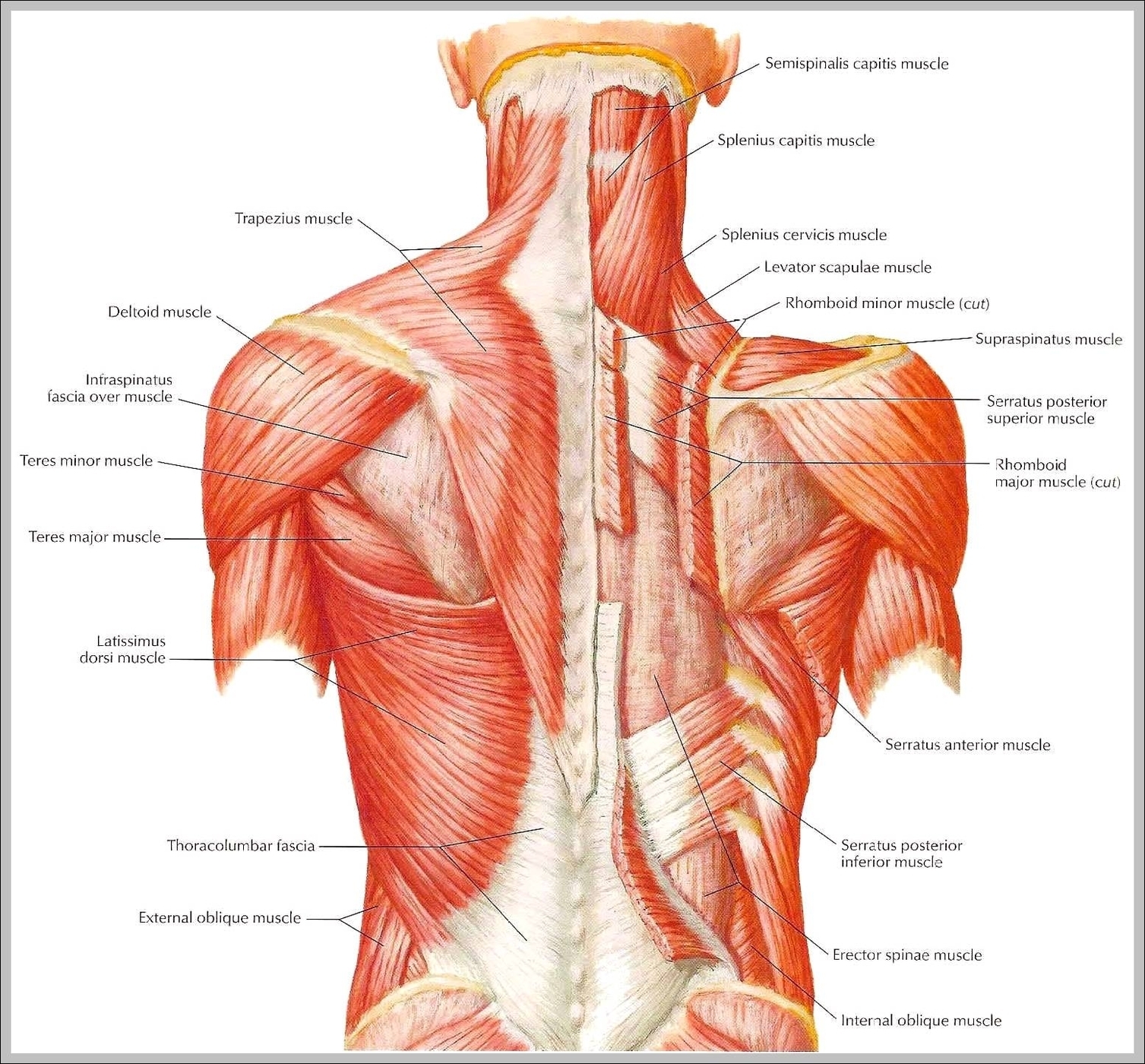
The upper body comprises seven general muscle regions in the shoulders, chest and back. Each of these regions contains several muscle groups consisting of several muscle heads, or parts.
The main muscle group of the chest is the pectorals. A well-developed chest is very important because it will add major size to your inner upper body. The chest is a very visible part of the upper body, that can add weight and force to all your martial arts moves. The pectorals or pecs are the large chest muscles.
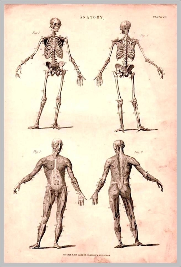
Upper Body Muscle Names Diagram - Chart - diagrams and charts with labels. This diagram depicts Upper Body Muscle Names