Hand Bones Labeled Image

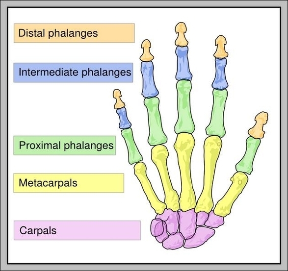
1,317 hand bone stock photos and images available, or search for skeleton hand or human bone to find more great stock photos and pictures. Carpal, metacarpal and phalanges of the hand, human body, drawing.

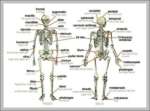
The wrist bones connect to the hand’s metacarpal bones. These are the largest bones of the hand. The ends of these five bones touch the wrist and create the skeletal structure for the palm. The metacarpals are numbered one through five. The thumb is number one and the pinky is number five.

148,045 hand anatomy stock photos, vectors, and illustrations are available royalty-free.

Hand Bones Labeled Image Diagram - Chart - diagrams and charts with labels. This diagram depicts Hand Bones Labeled Image

Hand Bones Labeled Image

Labeled Skeleton Diagram Image

62 labeled diagram of the skeletal system pic stock photos and images available, or start a new search to explore more stock photos and images. Skeletal Poster Human skeletal system poster containing detailed information about the skeletal structure.

Blank skeleton diagram to label front and back of the outstanding skeletal system anatomy human skeletal system human skeleton labeled. These workbooks are ideal for both youngsters and grown ups to utilize. Anatomy of the eye provide the labels for the indicated parts on the diagram of an eye.

The vertebrae of the dog skeleton anatomy also possess some exceptional osteological features that a ruminant or horse. Now, you should try to identify all the bones from the dog skeleton from the actual samples of your anatomy laboratory. You may use the dog bones labeled diagram from the anatomy learner.

Labeled Skeleton Diagram Image Diagram - Chart - diagrams and charts with labels. This diagram depicts Labeled Skeleton Diagram Image

Labeled Skeleton Diagram Image

Labeled Picture Of The Digestive System Image

125,099 human digestive system stock photos and images available, or search for human digestive system illustration or human digestive system anatomy to find more great stock photos and pictures.

In this system, the process of digestion has The human digestive system consists of the gastrointestinal tract plus the accessory organs of digestion.

The photo of large intestine is on the woman `s body, isolate on white background, Female anatomy concept Internal organs. The photo of internal organs is on the women`s body against gray background, Viscera on Human Woman using digital x-ray of human intestine holographic scan projection 3D rendering.

Labeled Picture Of The Digestive System Image Diagram - Chart - diagrams and charts with labels. This diagram depicts Labeled Picture Of The Digestive System Image

Labeled Picture Of The Digestive System Image

Picture Of Heart Labeled Image

7,463 human heart anatomy stock photos and images available, or search for organs or artery to find more great stock photos and pictures.

We will end with an exercise using a blank model of the heart, so you can quiz yourself on the gross anatomy by labeling and matching the main cardiac structures. By the end of this blog, you will be able to label a drawing of an anterior view (frontal section) of the heart as shown below.

Human Heart (Anatomy): Diagram, Function, Chambers, Location in Body WebMD’s Heart Anatomy Page provides a detailed image of the heart and provides information on heart conditions, tests, and treatments.

Picture Of Heart Labeled Image Diagram - Chart - diagrams and charts with labels. This diagram depicts Picture Of Heart Labeled Image

Picture Of Heart Labeled Image

Labeled Heart Image

Label the heart. In this interactive, you can label parts of the human heart. Drag and drop the text labels onto the boxes next to the diagram. Selecting or hovering over a box will highlight each area in the diagram. In this interactive, you can label parts of the human heart. Drag and drop the text labels onto the boxes next to the heart diagram.

199 heart labeled illustrations & vectors are available royalty-free. Fragile heart. Broken heart labeled fragile, fun illustration Congestive heart failure vector illustration. Labeled medical compare scheme. Congestive heart failure vector illustration. Labeled medical problem vs healthy Heart blood flow vector illustration.

We will end with an exercise using a blank model of the heart, so you can quiz yourself on the gross anatomy by labeling and matching the main cardiac structures. By the end of this blog, you will be able to label a drawing of an anterior view (frontal section) of the heart as shown below.

Labeled Heart Image Diagram - Chart - diagrams and charts with labels. This diagram depicts Labeled Heart Image

Labeled Heart Image

Diagram Blood Circulation Labeled Image

What is a Circulatory System Diagram. Systemic Circulation: After receiving oxygenated blood from the lungs the arteries of the systemic circulation system take the oxygenated blood from the heart to the rest of the body. The veins of the systemic circulation system take the then deoxygenated blood from the body back to the heart.

The veins of the systemic circulation system take the then deoxygenated blood from the body back to the heart. There are several different circulatory system diagrams. They may come with or without labels.

The blue arrows represent the flow of deoxygenated blood through the right side of the heart. The red arrows represent the flow of oxygenated blood through the left side of the heart. Diagram: Blue arrows demonstrate flow of deoxygenated blood through the right side of the heart.

Diagram Blood Circulation Labeled Image Diagram - Chart - diagrams and charts with labels. This diagram depicts Diagram Blood Circulation Labeled Image

Diagram Blood Circulation Labeled Image

Labeled Diagram Of Human Heart Image

Human Heart Diagram Labeled. The human heart is an organ responsible for pumping blood through the body, moving the blood (which carries valuable oxygen) to all the tissues in the body. Without the heart, the tissues couldn’t get the oxygen they need and would die.

Anatomy of the human heart as a focus and magnification of the circulation and cardiovascular system from a healthy body as a medical health care symbol of an inner vascular organ as a medical diagram. Section through the heart of human embryo showing the formation of the cardiac septa and the auriculo-ventricular valves, 5 to 6 weeks.

Human Heart (Anatomy): Diagram, Function, Chambers, Location in Body WebMD’s Heart Anatomy Page provides a detailed image of the heart and provides information on heart conditions, tests, and treatments.

Labeled Diagram Of Human Heart Image Diagram - Chart - diagrams and charts with labels. This diagram depicts Labeled Diagram Of Human Heart Image

Labeled Diagram Of Human Heart Image

Vertebrae Labeled Image

19,283 human vertebra stock photos and images available, or search for spine or human spine to find more great stock photos and pictures. section of the human vertebral column and cross-section of spinal cord. section of the human vertebral column and cross-section of spinal cord.

Vertebrae are boneslocated within the vertebral column. In humans, they are a series of 33 bonesthat run from the base of the skull to the coccyx. The irregularly shaped bones form the roughly S-shape of the spinal cord.

The spine is a darker white than the background, and shows all of the intricate details of the various parts from the top to the bottom. The spine is curved and shows spaces in between the vertebrae. human vertebra stock pictures, royalty-free photos & images The human spine on a white background.

Vertebrae Labeled Image Diagram - Chart - diagrams and charts with labels. This diagram depicts Vertebrae Labeled Image

Vertebrae Labeled Image

Skeletal System Labeled Image

63 labeled diagram of the skeletal system pics stock photos and images available, or start a new search to explore more stock photos and images. Carpal tunnel vector illustration scheme.

Save 25% when you use code SHARE25. 10,748 human skeleton labeled stock photos, vectors, and illustrations are available royalty-free.

Last Updated: Jul 29, 2020 The skeletal system includes all of the bones and joints in the body. Each bone is a complex living organ that is made up of many cells, protein fibers, and minerals. The skeleton acts as a scaffold by providing support and protection for the soft tissues that make up the rest of the body.

Skeletal System Labeled Image Diagram - Chart - diagrams and charts with labels. This diagram depicts Skeletal System Labeled Image

Skeletal System Labeled Image

Labeled Human Skeleton Diagram Image

2,854 human skeleton diagram stock photos and images available, or start a new search to explore more stock photos and images. human anatomy skeleton and muscles of the body – human skeleton diagram stock illustrations

Labeled Skeleton Diagram Cranium. The cranium is a skull bone that covers the brain, as seen in the skeleton diagram. The facial bones are not a… Stapes. Stapes is the smallest and the lightest bone in the human body. It is a stirrup-shaped bone found in the middle… Facial Bones. There are 12 …

First, divide the labeled diagram of skeleton in 4 sections, like above, which are head, arms, chest + hips and the fourth one – legs. In a similar manner use the method, for chest + hips section and hands section.

Labeled Human Skeleton Diagram Image Diagram - Chart - diagrams and charts with labels. This diagram depicts Labeled Human Skeleton Diagram Image

Labeled Human Skeleton Diagram Image

Labeled Muscular System Diagram Image

98 muscular system labeled diagram stock photos and images available, or start a new search to explore more stock photos and images. Frontal view of the muscular system of the male human body with descriptive labels pointing to the muscles on a white background. Labeled Anatomy Chart of Neck and Shoulder Muscles on White…

This muscular system picture shows all the major muscle groups on the human body from the frontal view. To see a muscular system diagram from the posterior (back) view click here.

Labeled human anatomy diagram of man’s full body muscular system from a posterior view on a white background. Labeled chart of the facial muscles of a woman, frontal view on a white background. Male Shoulder Ligaments and Biceps Muscles Isolated in Skeleton…

Labeled Muscular System Diagram Image Diagram - Chart - diagrams and charts with labels. This diagram depicts Labeled Muscular System Diagram Image

Labeled Muscular System Diagram Image

Labeled Diagram Of Digestive System Image

10,975 diagram of digestive system stock photos, vectors, and illustrations are available royalty-free. See diagram of digestive system stock video clips

The diagram of the human digestive system is useful for both Class 10 and 12. It is one among the few important topics, which are repetitively asked in the board examinations. The diagram below shows the structure and functions of the human digestive system.

Vector illustration of human digestive system Vector isolated illustration of human digestive system anatomy. Esophagus, stomach, duodenum, pancreas, intestine, gallbladder, liver icon. Medical information poster. Internal organ symbol design. digestive system diagram stock illustrations

Labeled Diagram Of Digestive System Image Diagram - Chart - diagrams and charts with labels. This diagram depicts Labeled Diagram Of Digestive System Image

Labeled Diagram Of Digestive System Image

Muscle Labeled Diagram Image

98 muscular system labeled diagram stock photos and images available, or start a new search to explore more stock photos and images. Frontal view of the muscular system of the male human body with descriptive labels pointing to the muscles on a white background. Labeled Anatomy Chart of Neck and Shoulder Muscles on White…

Medical labeled diagram with all kind cells. Example of blood, neurons, cardiac, bone, intestinal, epithelial, fat, liver and muscle. Muscle membrane vector illustration. Labeled scheme with myofibril, disc, zone, line and band.

Tibialis Anterior Muscle Tibialis Posterior Muscle Vastus Lateralis Muscle Vastus Medialis Muscle GENERAL Cross-Sections of Muscles of the Arm and Leg Muscle Cell Types Pronation/Supination Push/Pull Muscles Change Current View Angle Muscular System (Male View) Muscular System (Male Posterior View) Muscular System (Posterior View)

Muscle Labeled Diagram Image Diagram - Chart - diagrams and charts with labels. This diagram depicts Muscle Labeled Diagram Image

Muscle Labeled Diagram Image