Picture Of Neck Muscles

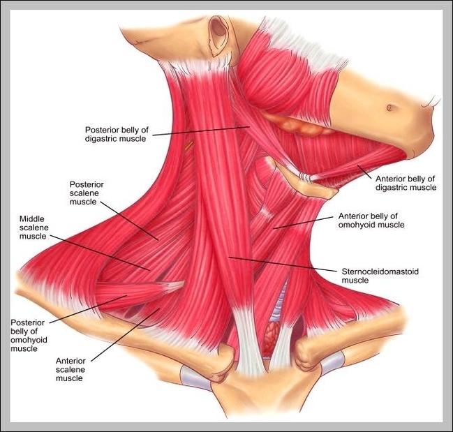
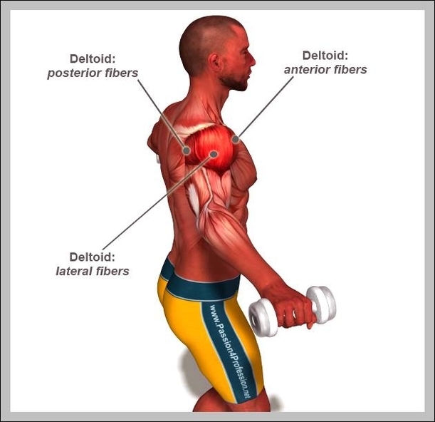
Muscles of neck. The muscles of the neck run from the base of the skull to the upper back and work together to bend the head and assist in breathing. The motion of the muscles of the neck are divided into four categories: rotation, lateral flexion, flexion, and hyperextension. Rotation describes the action of moving the head from side to side,…

These muscles and the deep muscles of the neck can be the causes of neck pain due to muscle strains, muscle tension, and other issues. A common cause of neck pain is muscle strain. This is often a result of incorrect posture.

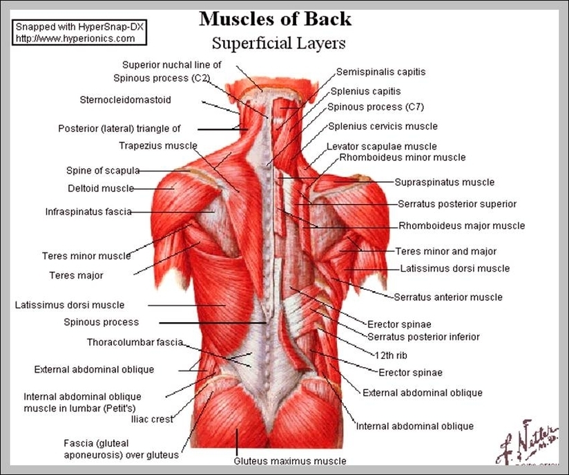
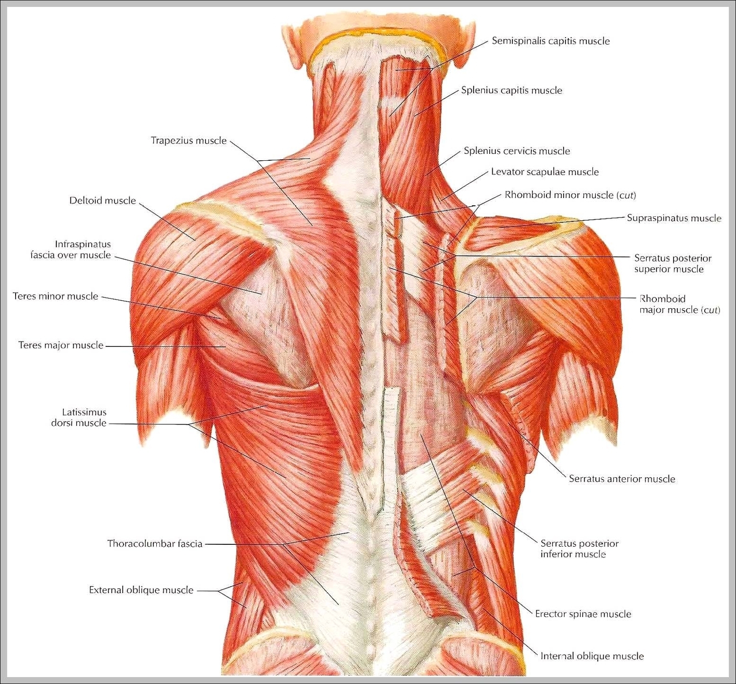
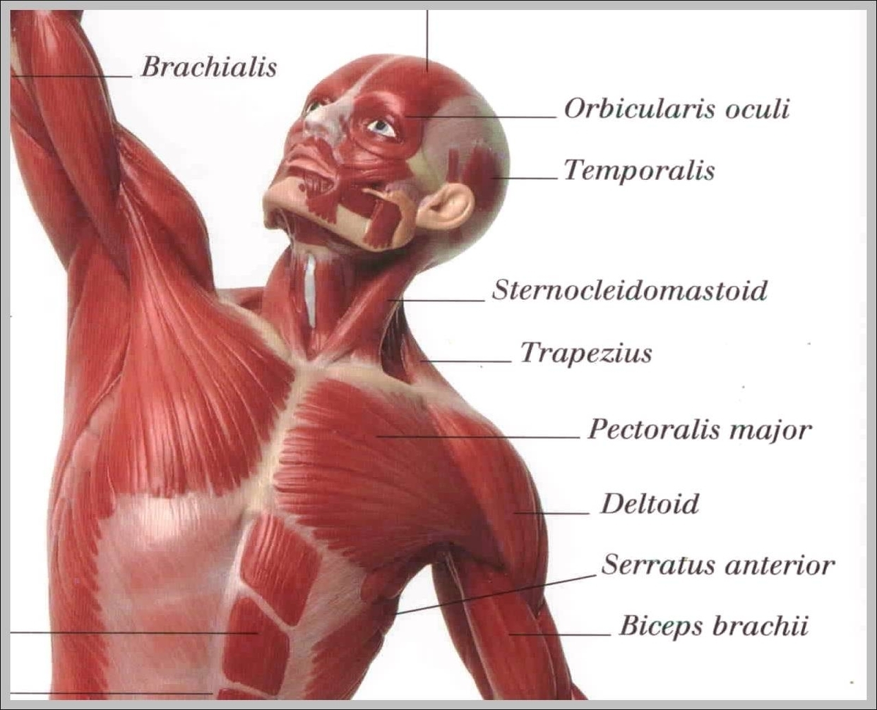
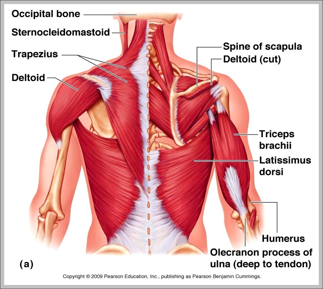
Superficial Muscles. Located underneath the platysma on the sides of the neck are the sternocleidomastoid muscles. With one on each side of the neck, these help flex the neck and rotate the head upward and side to side. They stretch from behind the ear diagonally to the center of the chest at the sternum.

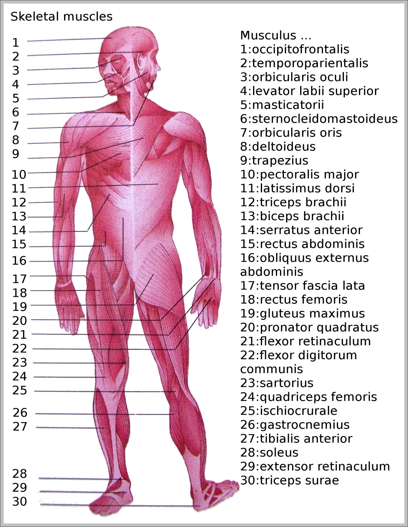
Picture Of Neck Muscles Diagram - Chart - diagrams and charts with labels. This diagram depicts Picture Of Neck Muscles

picture of neck muscles

What Are Muscle Cells

Types of muscle cells
Skeletal Muscle cells. Skeletal muscle cells are long, cylindrical, and striated.
Cardiac Muscle Cells. Cardiomyocytes are short and narrow, and fairly rectangular in shape.
Smooth Muscle Cells. Smooth muscle cells are spindle-shaped and contain a single central nucleus.

Muscle Cell Definition: A muscle cell is also known as Myocyte, which is a specialized animal cell that can shorten its length by using a series of motor proteins especially arranged in the cell.

The short, non-striated — and, hence — smooth muscle cells contain only one nucleus. Cardiac muscle cells appear striated, though they also appear less organized into stripes than skeletal muscle cells. These cells may branch out, forming physical connections with many surrounding cells.

What Are Muscle Cells Diagram - Chart - diagrams and charts with labels. This diagram depicts What Are Muscle Cells

what are muscle cells

Picture Of Human Leg Muscles

You may also find Vastus intermedius deep layer, Vastus medial, Adductor brevis m., Adductor longus m., Adductor Magnus & minimus m., Sartorius m., Gracilis m., Semimembranous muscle, Soleus m., Gastrocnemius m., Plantaris m. in it. We are pleased to provide you with the picture named Muscle of the human leg diagram.

The muscles that make up the quadriceps are the strongest and leanest of all muscles in the body. These four muscles at the front of the thigh are the major extensors (help to extend the leg …

In this image, you will find Muscle of the human leg diagram, Hip and femur middle layer, Hip and femur deep layer, Overview of the most important muscles of the leg, Femur middle layer, Femur deep layer, Rectus femoris m. in it.

Picture Of Human Leg Muscles Diagram - Chart - diagrams and charts with labels. This diagram depicts Picture Of Human Leg Muscles

picture of human leg muscles

Upper Back Muscle Diagram

Common causes of upper back pain 1. Muscle deconditioning and poor posture. Using the muscles infrequently or incorrectly can cause back pain. People can… 2. Muscle overuse. Overusing back muscles is another common cause of upper back pain. This typically occurs due to… 3. Traumatic injury. A …

Upper Back Muscles. The deltoid, teres major, teres minor, infraspinatus, supraspinatus (not shown) and subscapularis muscles (not shown) all extend from the scapula to the humerus and act on the shoulder joint. The trapezius and latissimus dorsi muscles connect the upper limb to the vertebral column.

Upper Back Muscles. The deltoid, teres major, teres minor, infraspinatus, supraspinatus (not shown) and subscapularis muscles (not shown) all extend from the scapula to the humerus and act on the shoulder joint.

Upper Back Muscle Diagram Diagram - Chart - diagrams and charts with labels. This diagram depicts Upper Back Muscle Diagram

upper back muscle diagram

Picture Of Muscles In Back

The back anatomy includes the latissimus dorsi, trapezius, erector spinae, rhomboid, and the teres major. On this page, you’ll learn about each of these muscles, their locations and functional anatomy. Function of the Back Muscles There are several individual muscles within the back anatomy, and it’s important to take a quick look at all of

Function of the Back Muscles. The back muscles enable you to stand up straight; support and protect your spine; and reach, pull and extend your arms and torso. Poorly developed back muscles lead to everything from muscle tweaks and pulls to imbalances of the musculature to the all-too-common hunched-over look (the “Neanderthal look”).

Back muscles are divided into two specific groups: the extrinsic muscles that are associated with upper extremity and shoulder movement, and the intrinsic muscles that deal with movements of the vertebral column. Several small muscles in the cervical area of the vertebral column are also important.

Picture Of Muscles In Back Diagram - Chart - diagrams and charts with labels. This diagram depicts Picture Of Muscles In Back

picture of muscles in back

Picture Of Human Skeleton With Muscles

There are around 640 skeletal muscles within the typical human body.

There are around 640 skeletal muscles within the typical human body. Almost every muscle constitutes one part of a pair of identical bilateral muscles, found on both sides, resulting in approximately 320 pairs of muscles, as presented in this article.

Attached to the bones of the skeletal system are about 700 named muscles that make up roughly half of a person’s body weight. Each of these muscles is a discrete organ constructed of skeletal muscle tissue, blood vessels, tendons, and nerves.

Picture Of Human Skeleton With Muscles Diagram - Chart - diagrams and charts with labels. This diagram depicts Picture Of Human Skeleton With Muscles

picture of human skeleton with muscles

Types Of Muscles

There are three types of muscle found in the human body: Skeletal Muscle. Cardiac Muscle (heart muscle) Skeletal muscle. Skeletal Muscles are those which attach to bones and have the main function of contracting to facilitate movement of our skeletons. They are also sometimes known as striated muscles due to their appearance. The cause of this …

There are three types of muscles: Skeletal muscles are attached mainly to the skeletal bones but some are also attached to other structures (such as the eyes for eye movement) and causes movements of the body. Cardiac muscle is responsible for the rhythmic contractions of the heart. Smooth muscle lines the walls of hollow organs.

The 3 types of muscle tissue are cardiac, smooth, and skeletal. Cardiac muscle cells are located in the walls of the heart, appear striated, and are under involuntary control.

Types Of Muscles Diagram - Chart - diagrams and charts with labels. This diagram depicts Types Of Muscles

types of muscles

Picture Of Deltoid Muscle

The deltoid muscles are shoulder muscles of the human body. These muscles are broken into three groups, which are the anterior, lateral, and posterior muscles. The anterior deltoid is the muscle located on the front area of the shoulder. This muscle is primarily used by the body to lift the arms straight overhead.

Located superior to the shoulder joint, the deltoid muscle works with the supraspinatus to abduct the arm at the shoulder. On the anterior side of the shoulder, the coracobrachialis, serratus anterior, pectoralis major, and pectoralis minor muscles work as a group to flex and adduct the scapula and humerus anteriorly toward the sternum.

It is named after the Greek letter delta, which is shaped like an equilateral triangle. The deltoid is attached by tendons to the skeleton at the clavicle (collarbone), scapula (shoulder blade), and humerus (upper arm bone).

Picture Of Deltoid Muscle Diagram - Chart - diagrams and charts with labels. This diagram depicts Picture Of Deltoid Muscle

picture of deltoid muscle

Skeletal Muscle Picture

Skeletal muscles are the muscles that we can see and feel through our skin. An individual skeletal muscle is considered an organ within the muscular system of the body. The skeletal muscle works with nerve tissue, connective tissue and vascular or blood tissue.

This is a table of skeletal muscles of the human anatomy. There are around 640 skeletal muscles within the typical human body.

Skeletal muscle is striated muscle tissue that is attached to bones. Skeletal muscle attaches to the bones. At the macroscopic level, the skeletal muscles are composed of a variety of layers. Working out strengthens skeletal muscles, making them more visible.

Skeletal Muscle Picture Diagram - Chart - diagrams and charts with labels. This diagram depicts Skeletal Muscle Picture

skeletal muscle picture

Shoulder Muscle Names

The shoulder girdle muscles make the scapula (shoulder blade) move. Muscles of the shoulder joint include the subscapularis, latissimus dorsi, infraspinatus, teres minor, teres major, supraspinatus, deltoid, and pectoralis major (pec muscles). On this page:

For that reason, and because of the dexterity of the shoulder joint itself, the musculature of the shoulder is complex, ranging from massive prime mover muscles to finer stabilizer and fixator muscles. The shoulder (or humeroscapular) joint is formed by the articulation of the head of the humerus with the scapula.

Toggle Anatomy System. On the anterior side of the shoulder, the coracobrachialis, serratus anterior, pectoralis major, and pectoralis minor muscles work as a group to flex and adduct the scapula and humerus anteriorly toward the sternum. The latissimus dorsi and teres major on the posterior side extend and adduct the arm towards the vertebrae…

Shoulder Muscle Names Diagram - Chart - diagrams and charts with labels. This diagram depicts Shoulder Muscle Names

shoulder muscle names

Picture Of Back Muscles

Back Anatomy: All About the Back Muscles 1 Function of the Back Muscles 2 Latissimus Dorsi (Lats) 3 Trapezius (Traps) 4 Erector Spinae (Spinal Erectors) 5 Rhomboid 6 Teres Major

Lower Back and Superficial Muscles The muscles of the lower back help stabilize, rotate, flex, and extend the spinal column, which is a bony tower of 24 vertebrae that gives the body structure and houses the spinal cord. The spinal cord and its nerves are the means by which the body and brain communicate with one another.

The latter attaches from the nuchal ligament and C7-T11 vertebrae to the root of the spine of scapula. They are supplied by the dorsal scapular nerve. Both muscles act upon the scapulothoracic joint where they draw the scapula superomedially, rotate the glenoid cavity inferiorly and support the position of scapula.

Picture Of Back Muscles Diagram - Chart - diagrams and charts with labels. This diagram depicts Picture Of Back Muscles

picture of back muscles

Visceral Muscles

…the vertebrate musculature are the visceral musculature and the somatic musculature (the striated muscles of the body wall). Somatic musculature may be divided into appendicular, or limb, muscles and axial muscles. The axial muscles include the muscles of the tail, trunk, and eyeballs as well as a group of muscles…

Superiorly, visceral fascia includes the pharyngobasilar and pharyngobuccal fascia and, as such, fuses to the cranial base surrounding the attachments of the superior constrictor muscles (Last 1978). Cervical visceral fascia extends inferiorly into the neck, surrounding the nasopharynx, oropharynx, and remaining cervical viscera.

…the vertebrate musculature are the visceral musculature and the somatic musculature (the striated muscles of the body wall). Somatic musculature may be divided into appendicular, or limb, muscles and axial muscles. The axial muscles include the muscles of the tail, trunk, and eyeballs as well as a group of muscles….

Visceral Muscles Diagram - Chart - diagrams and charts with labels. This diagram depicts Visceral Muscles

visceral muscles

Torso Muscle Diagram

Muscle Spasms in Torso: Causes and Treatments 1 Spasms felt throughout the chest and abdomen region can be caused by… 2 Pulled Muscle in the Chest. A pulled muscle in the chest can result in muscle spasms. 3 Injury to the Chest Wall. Injuries to the chest wall/muscle can actually be fairly common…

The human torso is also known as the ‘trunk’. It is the central part of the body, and it is from here that the neck and the limbs extend. Some of the most critical human body organs are situated within the torso. The upper part consists of the heart and the lungs; these are protected by the rib cage.

Anatomynote.com found Labelled Diagram Of The Muscles In The Human Body from plenty of anatomical pictures on the internet. We think this is the most useful anatomy picture that you need. You can click the image to magnify if you cannot see clearly. This image added by admin. Thank you for visit anatomynote.com.

Torso Muscle Diagram Diagram - Chart - diagrams and charts with labels. This diagram depicts Torso Muscle Diagram

torso muscle diagram

Visceral Smooth Muscle

Single-unit smooth muscle, or visceral smooth muscle is a type of smooth muscle found in the uterus, gastro-intestinal tract, and the bladder. In SUVSM, a single smooth muscle cell in a bundle is innervated by an autonomic nerve fiber.

Visceral muscle tissue, or smooth muscle, is tissue associated with the internal organs of the body, especially those in the abdominal cavity.

The key difference between multiunit and visceral smooth muscle lies in the way in which its individual cells function. In multiunit smooth muscle, the individual cells function independently, while in visceral smooth muscle, cells are dependent on each other for their function.

Visceral Smooth Muscle Diagram - Chart - diagrams and charts with labels. This diagram depicts Visceral Smooth Muscle

visceral smooth muscle

Upper Body Muscle

List of Upper Body Muscles
Neck and Shoulders. The neck primarily consists of the sternocleidomastoid and the splenius muscles,…
Chest. The chest region includes two muscle groups and one individual muscle.
Abdominals. The abdominals primarily consist of the rectus abdominis, an individual muscle,…
Arms. The arms comprise two muscle groups…

Numerous conditions can cause upper body muscle pain. Upper body muscle pain may occur on one or both sides of the body, and it may be temporary or long-lasting. Anatomically speaking, the upper body is composed of all the tissues and structures in the head, neck, torso and upper extremities.

Click on the muscle exercise to watch a video on how to properly do the exercise. Deltoids : The deltoids, also known as the shoulders contribute to both the front and back of the upper body. They are essential to building a strong upper body. Chest: The chest muscle is a major muscle in the upper body also.

Upper Body Muscle Diagram - Chart - diagrams and charts with labels. This diagram depicts Upper Body Muscle

upper body muscle

The Longest Muscle In The Body

The sartorius muscle (/sɑːrˈtɔːriəs/) is the longest muscle in the human body. It is a long, thin, superficial muscle that runs down the length of the thigh in the anterior compartment.

The sartorius muscle (/sɑːrˈtɔːriəs/) is the longest muscle in the human body. It is a long, thin, superficial muscle that runs down the length of the thigh in the anterior compartment.

The biggest muscle in the human body is the gluteus maximus, or the buttock muscles, also known as “the glutes.” These muscles (there is one on each side) help move the hips and thighs, and keep the trunk of the body upright.

The Longest Muscle In The Body Diagram - Chart - diagrams and charts with labels. This diagram depicts The Longest Muscle In The Body

the longest muscle in the body

Upper Body Muscle Anatomy

The major muscles in the upper torso of the body include: Trapezius: This muscle extends across the neck, shoulder, and back. Rhomboid major: Attached to the shoulder blade, this muscle is one of many that aids shoulder movement. Infraspinatus: This rotator cuff muscle helps raise and lower the arm.

The muscles of the chest and upper back occupy the thoracic region of the body inferior to the neck and superior to the abdominal region and include the muscles of the shoulders. These important muscles control many motions that involve moving the arms and head — such as throwing a ball, looking up at the sky, and raising your hand.

Muscle Anatomy: Human Anatomy Chart 1 Abs & Obliques Anatomy 2 Back Anatomy 3 Biceps Anatomy 4 Calves Anatomy 5 Chest Anatomy 6 Forearms Anatomy 7 Leg, Hip & Gluteal Anatomy 8 Neck Anatomy More …

Upper Body Muscle Anatomy Diagram - Chart - diagrams and charts with labels. This diagram depicts Upper Body Muscle Anatomy

upper body muscle anatomy

Skeletal Muscle Labeled

Skeletal Muscle Labeled Diagram - Chart - diagrams and charts with labels. This diagram depicts Skeletal Muscle Labeled

skeletal muscle labeled