Eye Diagram With Labels

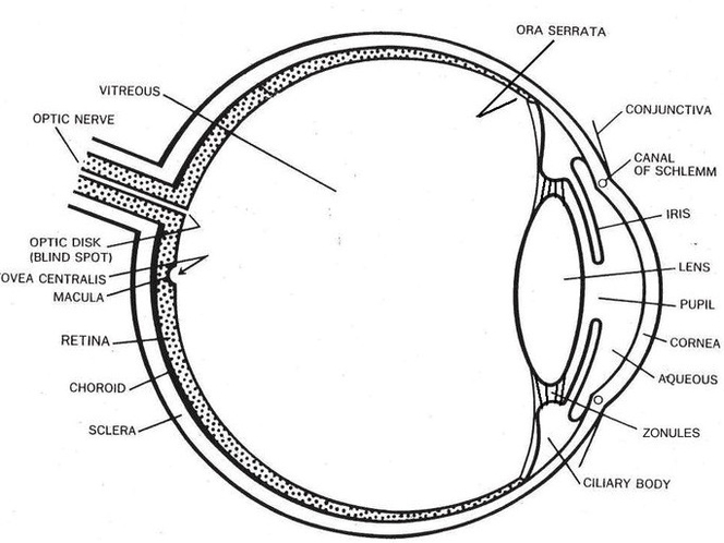
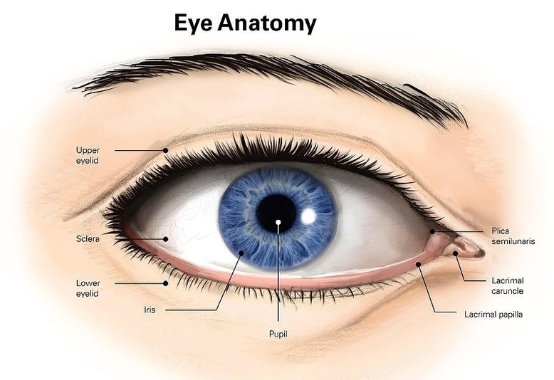
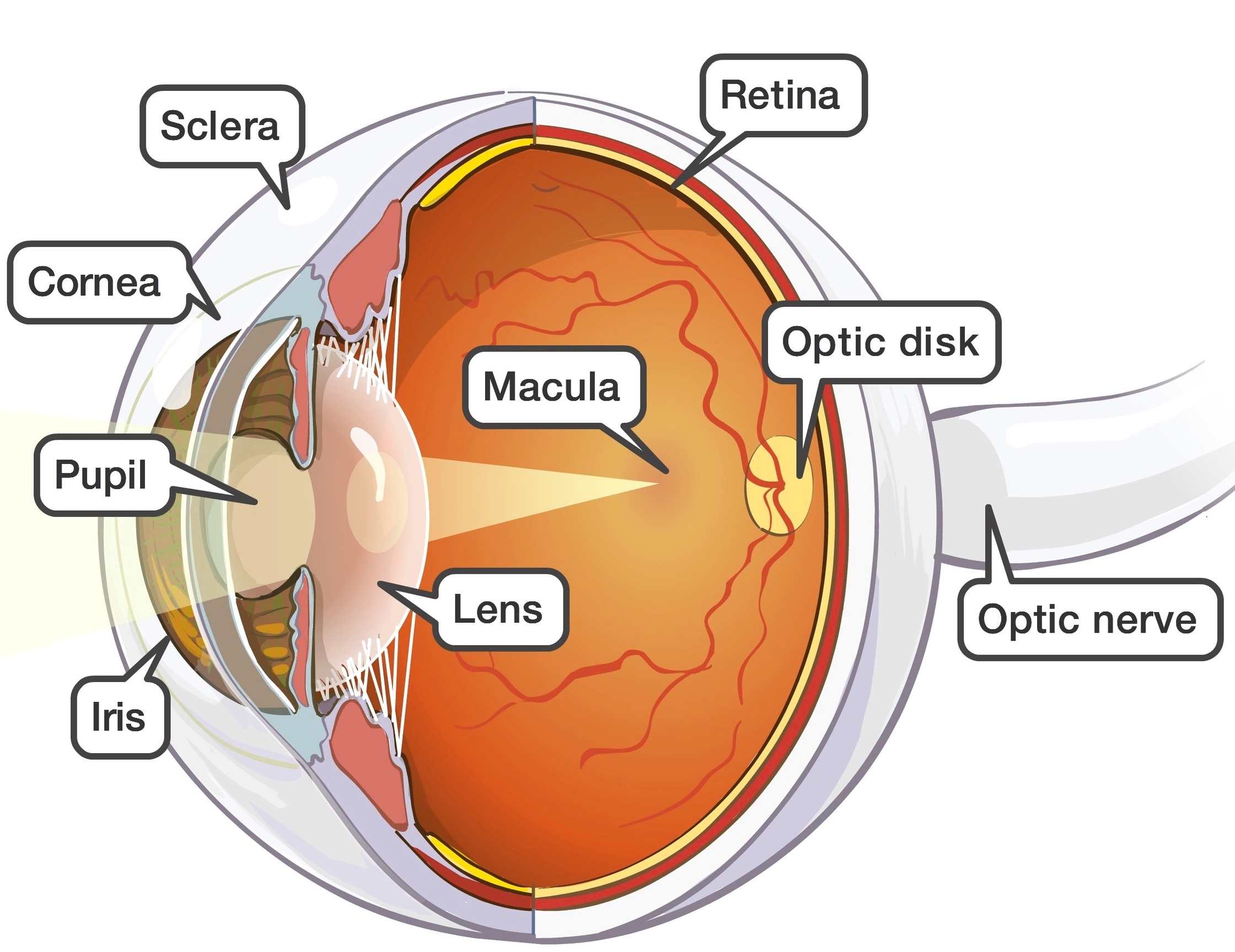
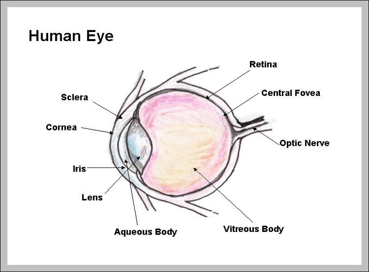
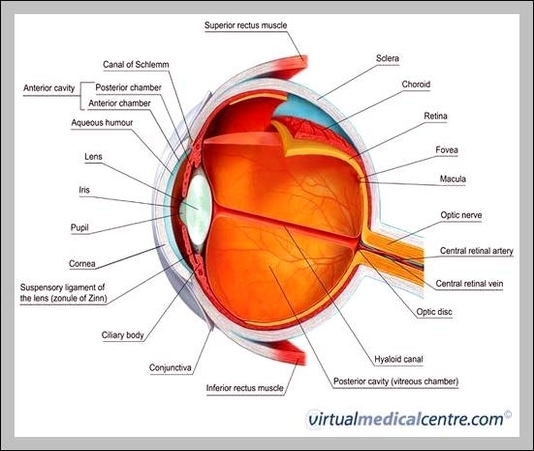
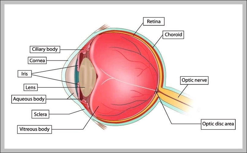
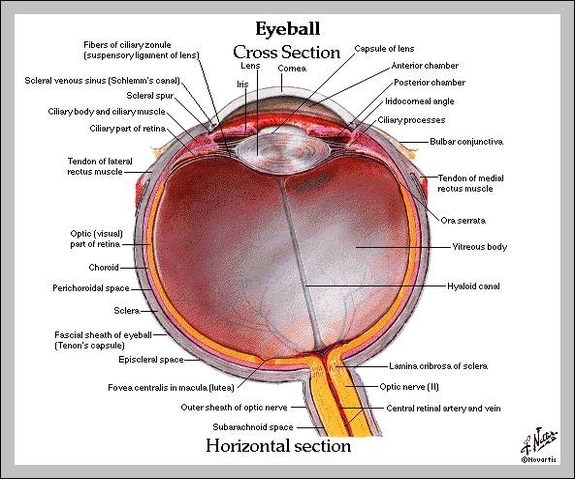
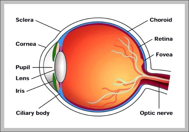
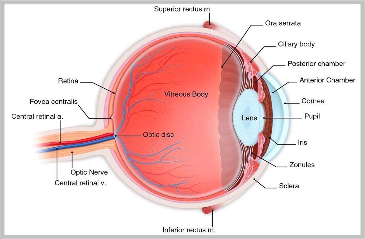
Use your mouse or finger to hover over a box to highlight the part to be named. Drag and drop the text labels onto the boxes next to the eye diagram If you want to redo an answer, click on the box and the answer will go back to the top so you can move it to another box.

Eye diagrams usually include voltage and time samples of the data acquired at some sample rate below the data rate. In Figure 1 , the bit sequences 011, 001, 100, and 110 are superimposed over one another to obtain the final eye diagram.

An eye diagram is a common indicator of the quality of signals in high-speed digital transmissions. An oscilloscope generates an eye diagram by overlaying sweeps of different segments of a long data stream driven by a master clock.

Eye Diagram With Labels Diagram - Chart - diagrams and charts with labels. This diagram depicts Eye Diagram With Labels

Eye diagram with labels

Eye Anatomy Both Internal And External Views

External and internal eye anatomy. Overview. The cornea allows light to enter the eye. As light passes through the eye the iris changes shape by expanding and letting more light through or constricting and letting less light through to change pupil size. The lens then changes shape to allow the accurate focusing of light on the retina.

Eye Structures (Front and Side Views) The slight bulge in the sclera at the front of the eye is a clear, thin, dome-shaped tissue called the cornea. The cornea directs light rays into the eye and helps focus them on the retina. The pupil is the opening in the coloured part of the eye (iris). It allows light to pass to the lens.

The external parts of the eye work together to protect the eye and all of its internal structures. The upper and lower eyelids form a moist region around the eye, and protect the surface of the eye from injury, infection, and disease. The eyelids contain muscles that enable them to open and close around the eye, and are covered with skin.

Eye Anatomy Both Internal And External Views Diagram - Chart - diagrams and charts with labels. This diagram depicts Eye Anatomy Both Internal And External Views

Eye anatomy both internal and external views

Eye Muscles Diagram

Eye muscles. There are two groups of eye muscles: Six extraocular muscles move the eye: superior rectus, inferior rectus, medial rectus, lateral rectus, superior oblique and inferior oblique muscles; and one other, levator palpebrae superioris, opens the eyelid.

Intrinsic ocular muscles which are within the eyeball itself and control how the eyes accommodate Six extraocular muscles move the eye: superior rectus, inferior rectus, medial rectus, lateral rectus, superior oblique and inferior oblique muscles; and one other, levator palpebrae superioris, opens the eyelid.

There are four recti muscles; superior rectus, inferior rectus, medial rectus and lateral rectus. These muscles characteristically originate from the common tendinous ring. This is a ring of fibrous tissue, which surrounds the optic canal at the back of the orbit.

Eye Muscles Diagram Diagram - Chart - diagrams and charts with labels. This diagram depicts Eye Muscles Diagram

Eye muscles diagram

Eye Section View

Eye – Cross Section. A human eyeball cross section showing the following structures: iris, anterior limiting membrane, posterior limiting membrane, tendon of Eye anatomy, Human eye cross section physiology, Model of cornea and lens for ophthalmologist. Eye anatomy, Human eye cross section physiology, Model of cornea Enlarged anatomical eye model.

The anatomy of the eye includes the cornea, pupil, lens, sclera, conjunctiva and more. There are many parts of the eye. My Dashboard My Education Find an Ophthalmologist

A section view provides architects and contractors another way of understanding how a construction will stand up, as it reveals details of the construction of walls and the thickness and height of beams and other supports.

Eye Section View Diagram - Chart - diagrams and charts with labels. This diagram depicts Eye Section View

Eye section view