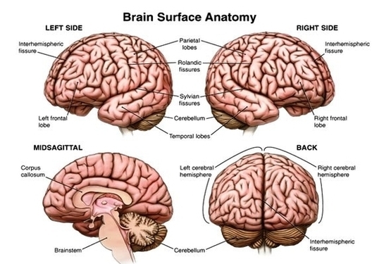
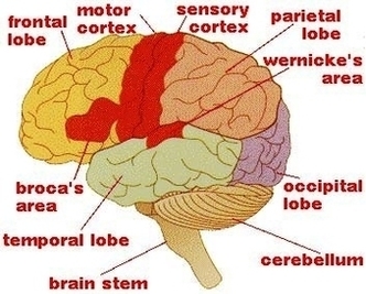
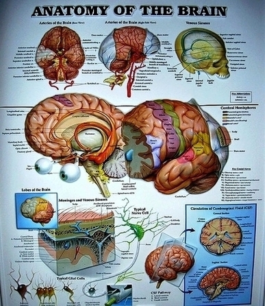
Before we have a look at the brain diagram, it is important to go through a few facts about the brain and its function. This will help you understand the anatomy of the brain better. The average dimension of the adult human brain is 5.5 inches in width and 6.5 inches in length.
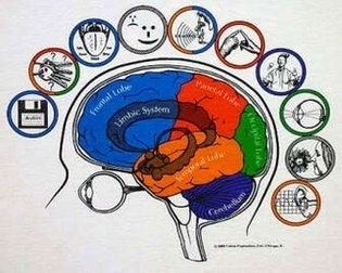
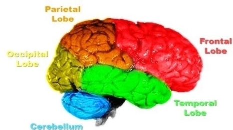
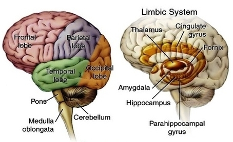
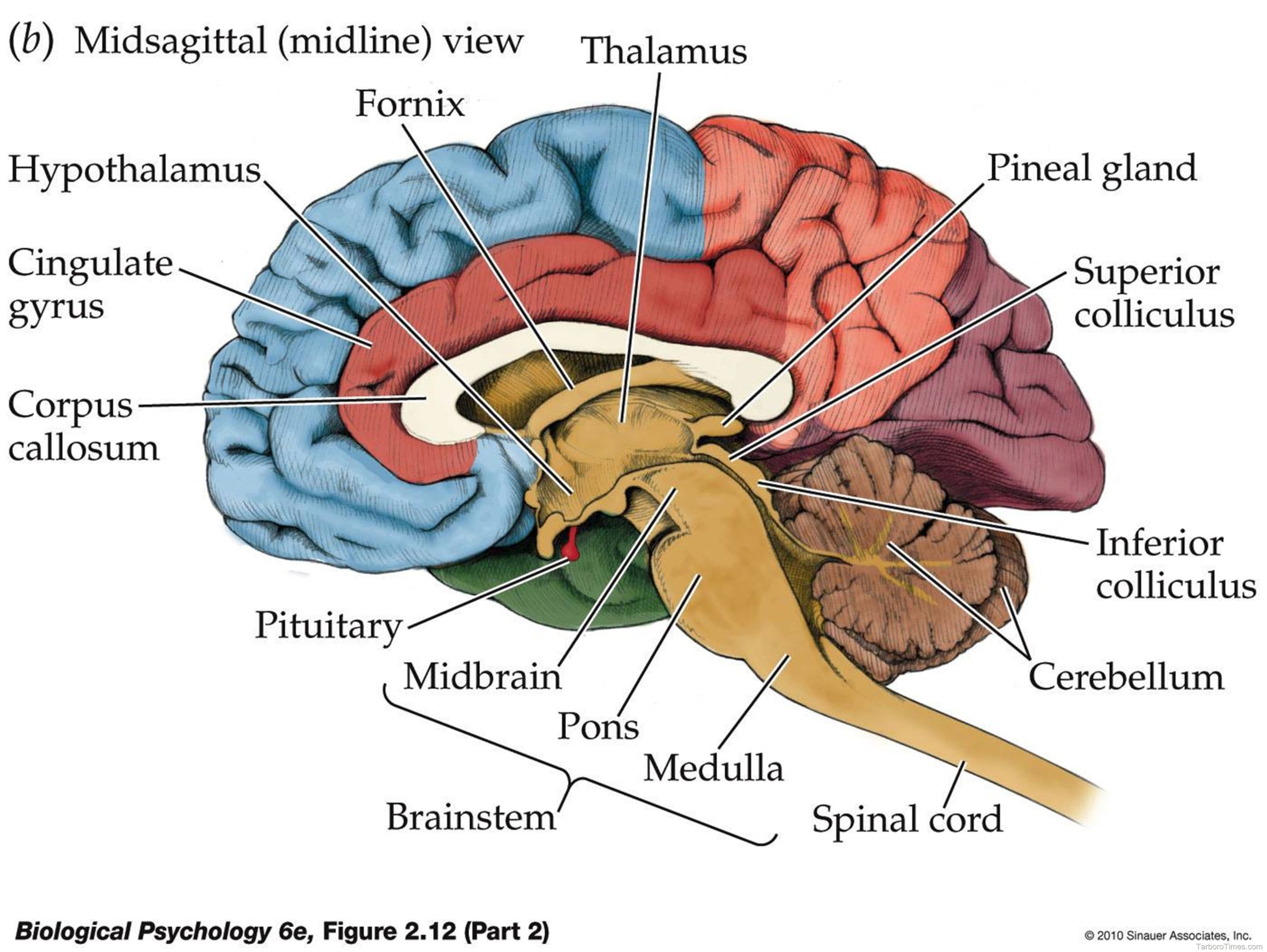
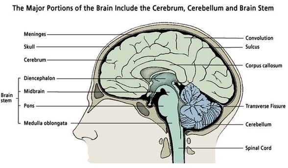
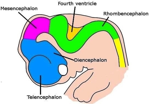
The diagram of the brain is useful for both Class 10 and 12. It is one among the few topics having the highest weightage of marks and is frequently asked in the examinations. A well-labelled diagram of a human brain is given below for further reference. The human brain is divided into three main parts:
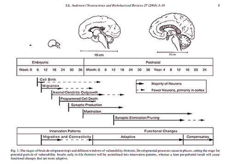
Let’s review each of the five stages of human brain growth: Stage 1: 0 to 10 months. Neurons and connections growing. Pregnant woman should stay as stress-free as possible, take folic acid, B6 & B12, stimulate this young developing brain with sounds and sensations. Mother should avoid toxins, cigarettes, heavy metals, alcohol, drugs.
Diagram Of Px Human Brain Development Timeline Image Diagram - Chart - diagrams and charts with labels. This diagram depicts Diagram Of Px Human Brain Development Timeline Image