Arm Bones Diagram Image

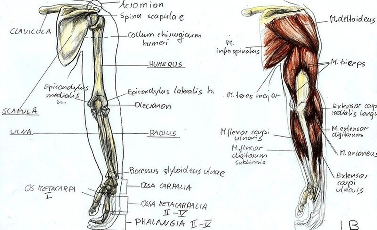
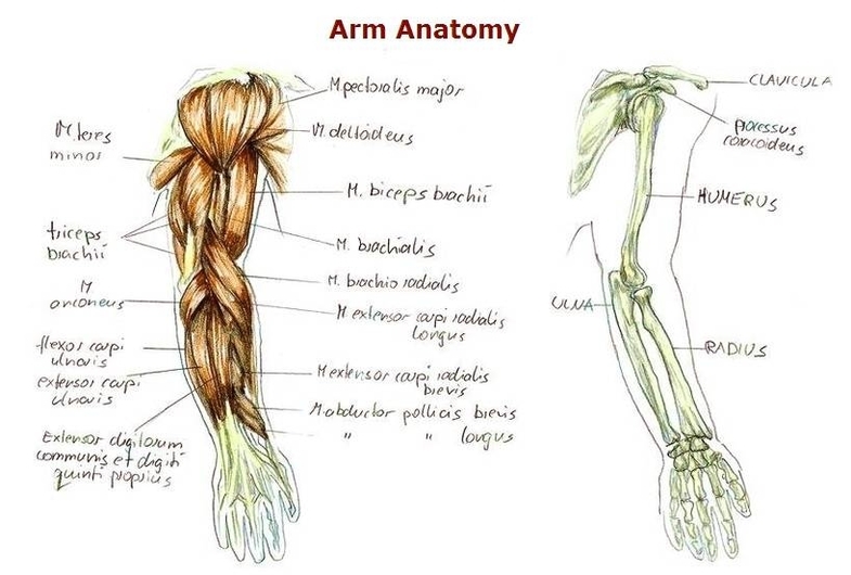
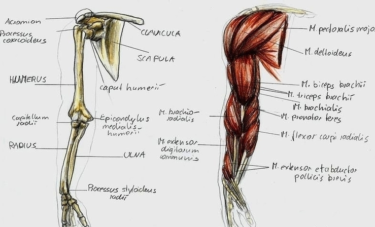
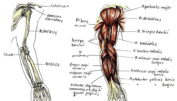
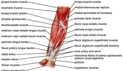
The human arm is divided into two main sections: the forearm and the arm itself. The forearm is the section located between the elbow and the wrist, while the arm represents the section from the shoulder to the elbow. The main bone of the upper arm is the humerus. By contrast, the forearm consists of two main bones, the radius and the ulna.

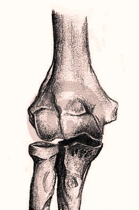
The large bones of the arm include: Humerus: This bone runs down from the shoulder socket and joins the radius and ulna at the elbow. Radius: A forearm bone, it runs from the elbow to the thumb side of the wrist. Ulna: This forearm bone runs from the elbow to the “pinkie” side of the wrist. These three bones join to form the elbow.

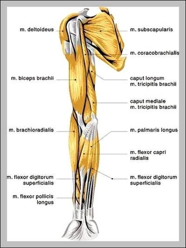
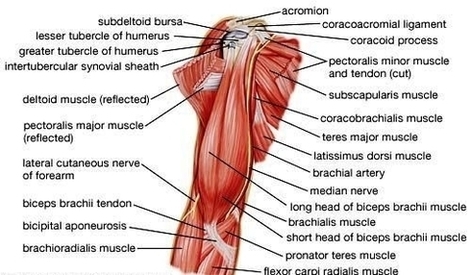
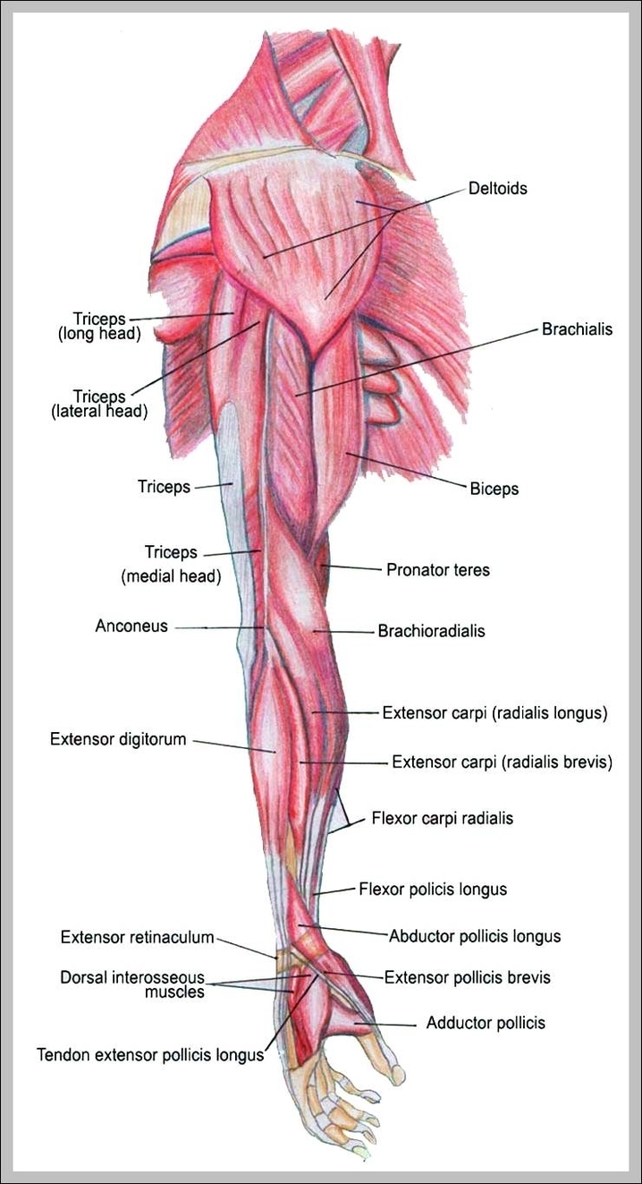
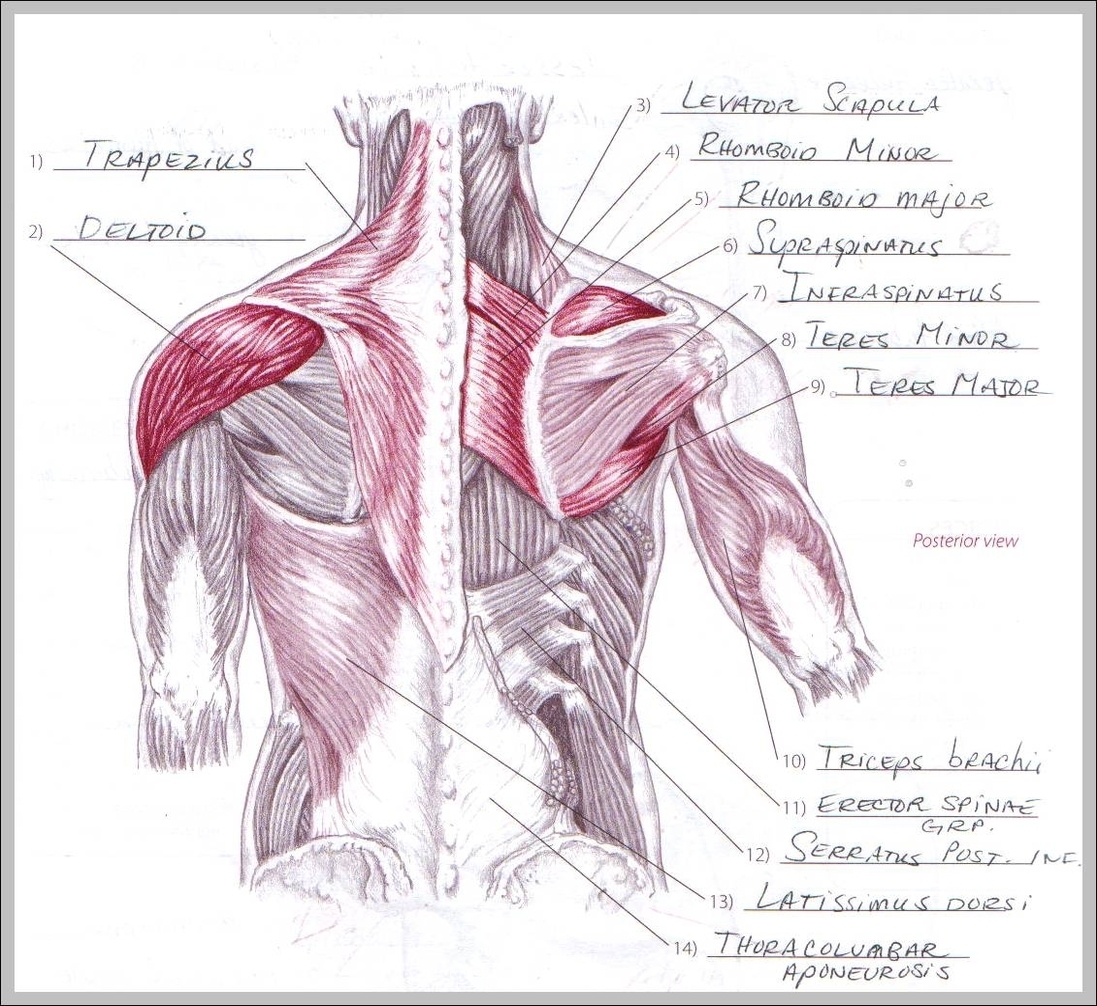
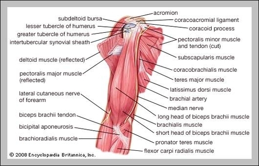
The bones of the arm and hand have the important jobs of supporting the upper limb and providing attachment points for the muscles that move the upper limb.

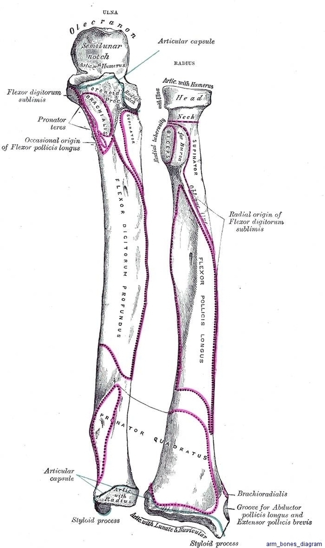
Arm Bones Diagram Image Diagram - Chart - diagrams and charts with labels. This diagram depicts Arm Bones Diagram Image

Arm Bones Diagram Image

Anatomy Arm By Bk Image

58,682 arm anatomy stock photos, vectors, and illustrations are available royalty-free. See arm anatomy stock video clips

The human arm is divided into two main regions, the portion from the elbow to the wrist known as the forearm, and the segment from the shoulder to the elbow referred to as the arm. Arm muscle anatomy enables the arm to perform a variety of movements, including flexion, extension, pronation, and supination.

The (upper) arm muscles are a group of five muscles located in the region between the shoulder and elbow joints. They are divided into two distinct compartments of the arm.

Anatomy Arm By Bk Image Diagram - Chart - diagrams and charts with labels. This diagram depicts Anatomy Arm By Bk Image

Anatomy Arm By Bk Image

Arm Muscles Image

27,539 arm muscle anatomy stock photos, vectors, and illustrations are available royalty-free.

In this article, we shall look at the anatomy of the muscles of the upper arm – their attachments, innervation and actions. There are three muscles located in the anterior compartment of the upper arm – biceps brachii, coracobrachialis and brachialis.

Anterior Compartment 1 Biceps Brachii. The biceps brachii is a two-headed muscle. … 2 Coracobrachialis. The coracobrachialis muscle lies deep to the biceps brachii in the arm. … 3 Brachialis. The brachialis muscle lies deep to the biceps brachii, and is found more distally than the other muscles of the arm.

Arm Muscles Image Diagram - Chart - diagrams and charts with labels. This diagram depicts Arm Muscles Image

Arm Muscles Image

Diagram Of Human Anatomy Muscles Arm Tkdcw Image

The human arm is divided into two main regions, the portion from the elbow to the wrist known as the forearm, and the segment from the shoulder to the elbow referred to as the arm. Arm muscle anatomy enables the arm to perform a variety of movements, including flexion, extension, pronation, and supination.

105,188 human muscle anatomy stock photos, vectors, and illustrations are available royalty-free.

Each of these two sections of the human arm consists of arm muscle anatomy that allows for the flexion, extension, pronation, and supination of the arm, as will be further discussed below. While four muscles are responsible for the upper arm musculature, there are over twenty muscles that move the forearm, wrist, hands, and fingers.

Diagram Of Human Anatomy Muscles Arm Tkdcw Image Diagram - Chart - diagrams and charts with labels. This diagram depicts Diagram Of Human Anatomy Muscles Arm Tkdcw Image

Diagram Of Human Anatomy Muscles Arm Tkdcw Image

Arm Anatomy1 Image

4,995 human arm anatomy stock photos and images available, or start a new search to explore more stock photos and images.

The human arm is divided into two main regions, the portion from the elbow to the wrist known as the forearm, and the segment from the shoulder to the elbow referred to as the arm. Arm muscle anatomy enables the arm to perform a variety of movements, including flexion, extension, pronation, and supination.

The muscles of the arms attach to the shoulder blade, upper arm bone ( humerus ), forearm bones ( radius and ulna ), wrist, fingers, and thumbs. These muscles control movement at the elbow, forearm, wrist, and fingers. When affected by injury or neuromuscular disorders, everyday tasks that require hand and arm use can be challenging.

Arm Anatomy1 Image Diagram - Chart - diagrams and charts with labels. This diagram depicts Arm Anatomy1 Image

Arm Anatomy1 Image

Arm Muscles Image

27,539 arm muscle anatomy stock photos, vectors, and illustrations are available royalty-free.

In this article, we shall look at the anatomy of the muscles of the upper arm – their attachments, innervation and actions. There are three muscles located in the anterior compartment of the upper arm – biceps brachii, coracobrachialis and brachialis.

Anterior Compartment 1 Biceps Brachii. The biceps brachii is a two-headed muscle. … 2 Coracobrachialis. The coracobrachialis muscle lies deep to the biceps brachii in the arm. … 3 Brachialis. The brachialis muscle lies deep to the biceps brachii, and is found more distally than the other muscles of the arm.

Arm Muscles Image Diagram - Chart - diagrams and charts with labels. This diagram depicts Arm Muscles Image

Arm Muscles Image

Muscles Arm Human Anatomy Illustration Diagram En Medical Image

The human arm is divided into two main regions, the portion from the elbow to the wrist known as the forearm, and the segment from the shoulder to the elbow referred to as the arm. Arm muscle anatomy enables the arm to perform a variety of movements, including flexion, extension, pronation, and supination.

Each of these two sections of the human arm consists of arm muscle anatomy that allows for the flexion, extension, pronation, and supination of the arm, as will be further discussed below. While four muscles are responsible for the upper arm musculature, there are over twenty muscles that move the forearm, wrist, hands, and fingers.

Arm muscle anatomy enables the arm to perform a variety of movements, including flexion, extension, pronation, and supination. The forearm flexors include the biceps brachii, coracobrachialis, and brachialis muscles, which help to bend the arm at the elbow, as when flexing one’s arm to show off the muscles.

Muscles Arm Human Anatomy Illustration Diagram En Medical Image Diagram - Chart - diagrams and charts with labels. This diagram depicts Muscles Arm Human Anatomy Illustration Diagram En Medical Image

Muscles Arm Human Anatomy Illustration Diagram En Medical Image

Anatomy Arm Nerves Image

There are four main nerves of the arm and they wrap around the bones. The ulnar and median nerves originate in the brachial plexus, which is a bundle of nerve fibers extending from the spinal cord to the arm and ending in the hand.

Arm and shoulder anatomy 1 Glenohumeral joint. The glenohumeral joint is one of the joints associated with the shoulder girdle that allow a full range of movement of the upper limb . 2 Bones. The shoulder is the region where the upper limb is attached to the trunk. … 3 Muscles. … 4 Nerves. … 5 Arteries and veins. …

58,682 arm anatomy stock photos, vectors, and illustrations are available royalty-free. See arm anatomy stock video clips

Anatomy Arm Nerves Image Diagram - Chart - diagrams and charts with labels. This diagram depicts Anatomy Arm Nerves Image

Anatomy Arm Nerves Image