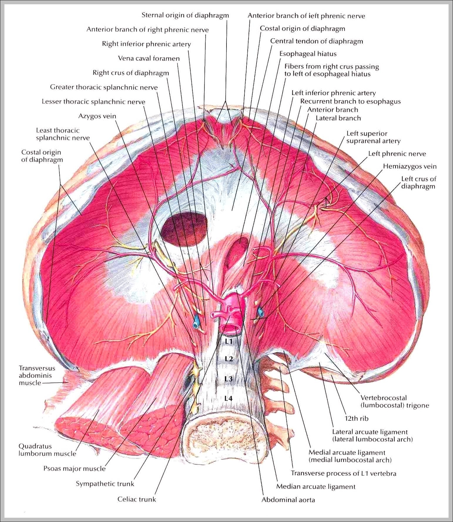
Diaphragm. The diaphragm is the dome-shaped sheet of muscle and tendon that serves as the main muscle of respiration and plays a vital role in the breathing process. Also known as the thoracic diaphragm, it serves as an important anatomical landmark that separates the thorax, or chest, from the abdomen.
The diaphragm has openings that allow certain structures to span the chest and abdominal cavities. As it moves rhythmically, the diaphragm remains anchored to the ribs, sternum (breastbone), and the spine.
The origins of the diaphragm are found along the lumbar vertebrae of the spine and the inferior border of the ribs and sternum. Openings in the diaphragm allow the esophagus, phrenic and vagus nerves, descending aorta, and inferior vena cava to pass between the thoracic and abdominal cavities.
Picture Of Diaphragm