Back Anatomy: All About the Back Muscles 1 Function of the Back Muscles 2 Latissimus Dorsi (Lats) 3 Trapezius (Traps) 4 Erector Spinae (Spinal Erectors) 5 Rhomboid 6 Teres Major
Lower Back and Superficial Muscles The muscles of the lower back help stabilize, rotate, flex, and extend the spinal column, which is a bony tower of 24 vertebrae that gives the body structure and houses the spinal cord. The spinal cord and its nerves are the means by which the body and brain communicate with one another.
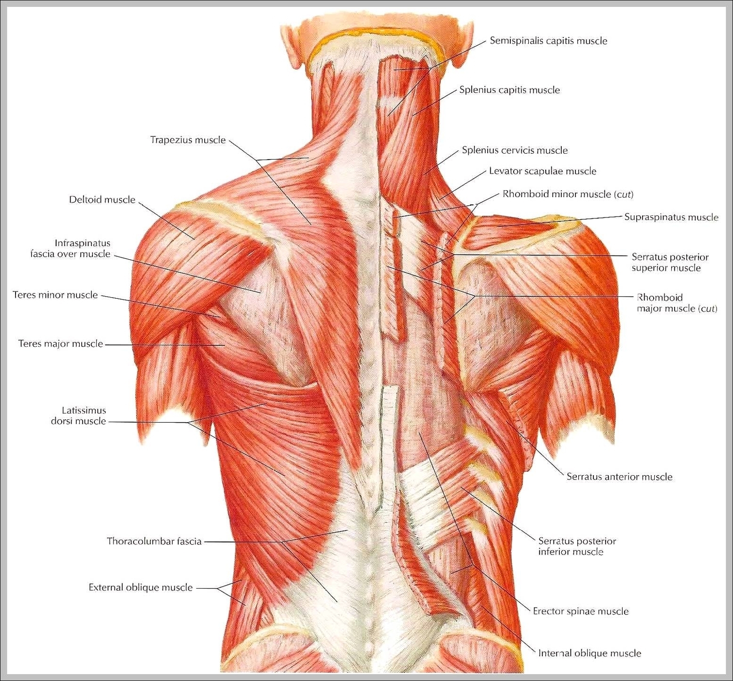
The latter attaches from the nuchal ligament and C7-T11 vertebrae to the root of the spine of scapula. They are supplied by the dorsal scapular nerve. Both muscles act upon the scapulothoracic joint where they draw the scapula superomedially, rotate the glenoid cavity inferiorly and support the position of scapula.
Picture Of Back Muscles