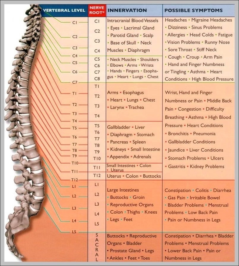
Definition of Body Systems We can define body systems as groups of organs and tissues that work together to perform important jobs for the body. There are some organs in our body which are part of more than one body system as they serve more than one function. Apart from these, other organs and tissues serve only one purpose in the body system.
The main systems of the human body are:
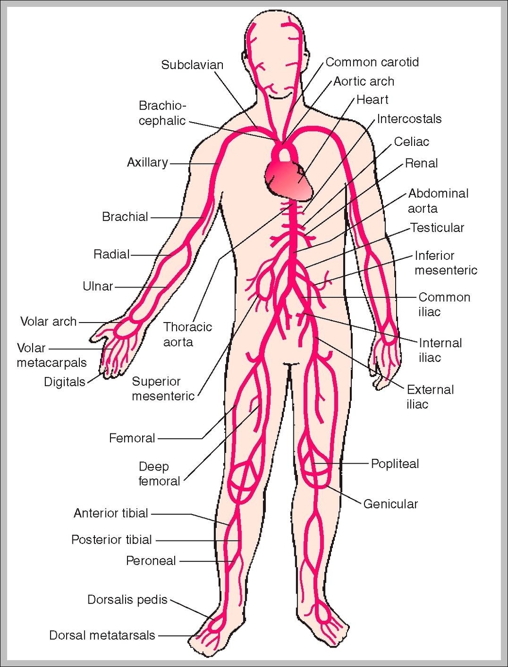
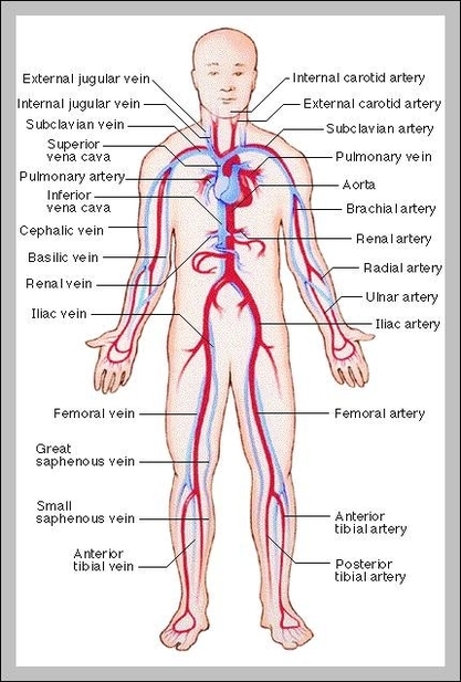
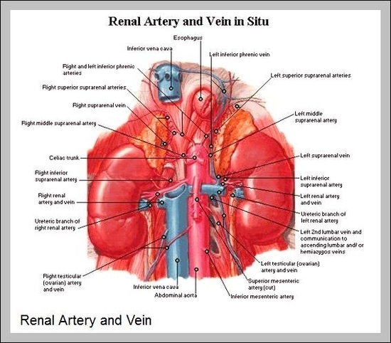
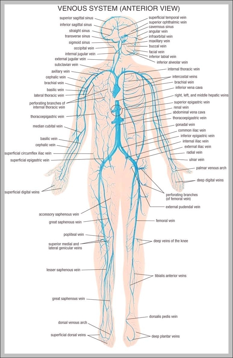
Circulatory system :
Circulates blood around the body via the heart, arteries and veins, delivering oxygen and nutrients to organs and cells and carrying their waste products away.
Keeps the body’s temperature in a safe range.
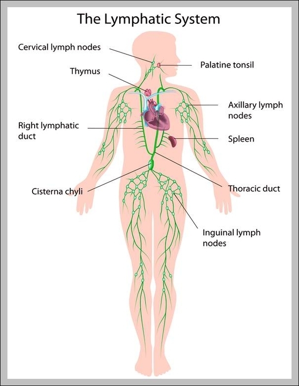
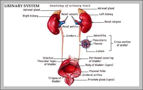
Digestive system and Excretory system :
More items…
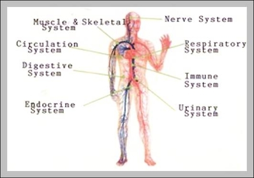
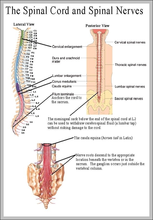
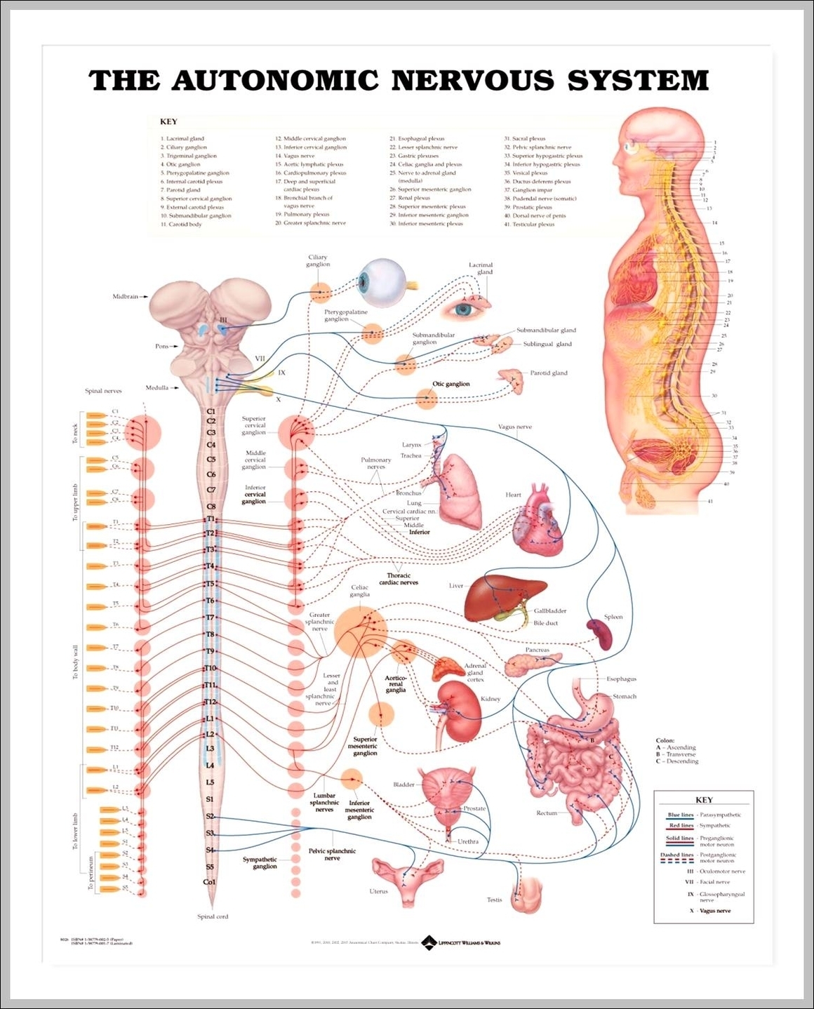
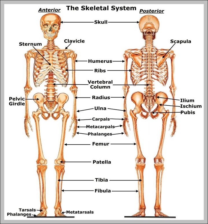
The highest level of body organization, then, is that of the organ system. The body includes nine major organ systems, each composed of various organs and tissues that work together as a functional unit. The chief constituents and prime functions of each system are summarized below.
System Of The Body Diagram - Chart - diagrams and charts with labels. This diagram depicts System Of The Body