Anatomy Of The Leg Muscles Image

72,510 leg anatomy stock photos, vectors, and illustrations are available royalty-free.

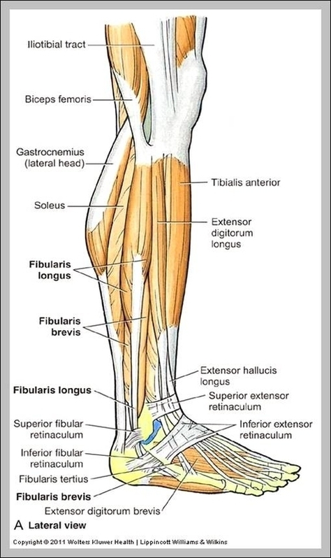
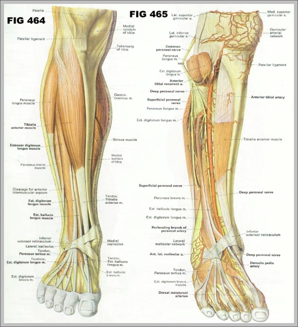
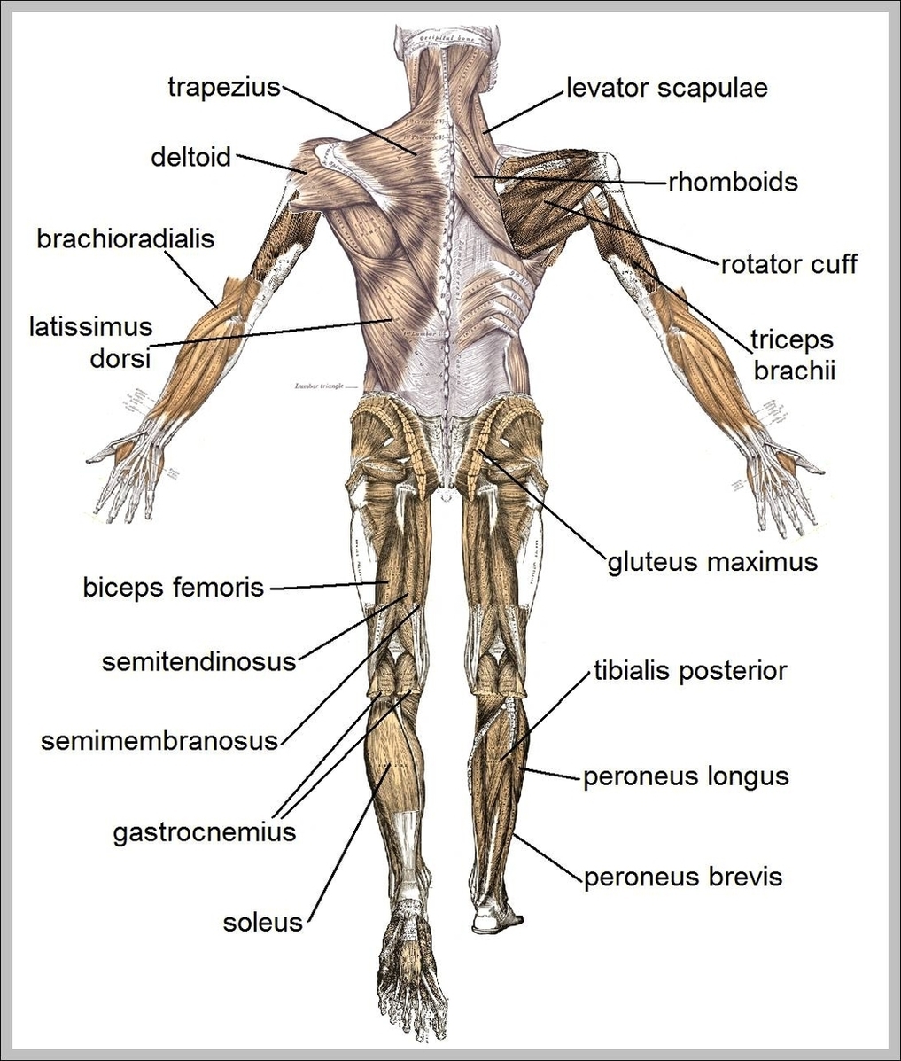
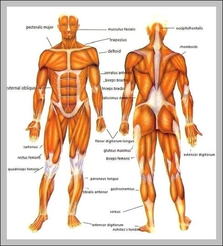
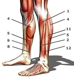
Leg muscles are bundles of fibrous tissue that contract and relax to exert forces on bones and move the legs The main muscle groups in the legs are: quadriceps, hamstrings, adductors in the upper leg or thigh, and the calves in the lower legs.

The legs are the lower extremities that allow for standing, walking, running, and more. Their movement is produced by the contraction and relaxation of leg muscles and their connection to the skeleton through tendons. The movement of the legs produced by these muscles happens around the leg joints, particularly the hips, knees and ankles.

Anatomy Of The Leg Muscles Image Diagram - Chart - diagrams and charts with labels. This diagram depicts Anatomy Of The Leg Muscles Image

Anatomy Of The Leg Muscles Image

Muscles Diagram Image

Last Updated: Jul 16, 2019 The muscular system is responsible for the movement of the human body. Attached to the bones of the skeletal system are about 700 named muscles that make up roughly half of a person’s body weight. Each of these muscles is a discrete organ constructed of skeletal muscle tissue, blood vessels, tendons, and nerves.

Tibialis Anterior Muscle Tibialis Posterior Muscle Vastus Lateralis Muscle Vastus Medialis Muscle GENERAL Cross-Sections of Muscles of the Arm and Leg Muscle Cell Types Pronation/Supination Push/Pull Muscles Change Current View Angle Muscular System (Male View) Muscular System (Male Posterior View) Muscular System (Posterior View)

Finally, the direction in which the muscle fibers run can be used to identify a muscle. In the abdominal region, there are several sets of wide, flat muscles.

Muscles Diagram Image Diagram - Chart - diagrams and charts with labels. This diagram depicts Muscles Diagram Image

Muscles Diagram Image

Human Muscle Structure Image

105,188 human muscle anatomy stock photos, vectors, and illustrations are available royalty-free.

Last Updated: Jul 16, 2019 The muscular system is responsible for the movement of the human body. Attached to the bones of the skeletal system are about 700 named muscles that make up roughly half of a person’s body weight. Each of these muscles is a discrete organ constructed of skeletal muscle tissue, blood vessels, tendons, and nerves.

Forearms – Anatomy Muscles Rhomboid minor and rhomboid major, levator scapulae and latissimus dorsi muscles – didactic board of anatomy of human bony and muscular system, posterior view Abs – Anatomy Muscles Leg muscles of the man Human Body Organs (Lungs) 3D Triceps – Anatomy Muscles Hand muscle connection with brain

Human Muscle Structure Image Diagram - Chart - diagrams and charts with labels. This diagram depicts Human Muscle Structure Image

Human Muscle Structure Image

Muscles In The Arms Image

27,539 arm muscle anatomy stock photos, vectors, and illustrations are available royalty-free.

What Are the Muscles of Your Arms? There are four main muscles of your arms: biceps, triceps, forearm flexors, and forearm extensors. There are also a handful of other muscles that support these main four.

The human arm is divided into two main regions, the portion from the elbow to the wrist known as the forearm, and the segment from the shoulder to the elbow referred to as the arm. Arm muscle anatomy enables the arm to perform a variety of movements, including flexion, extension, pronation, and supination.

Muscles In The Arms Image Diagram - Chart - diagrams and charts with labels. This diagram depicts Muscles In The Arms Image

Muscles In The Arms Image

Human Muscles Chart Image

Our human muscle anatomy charts shows all of the major muscle groups in a superficial, anterior view (that’s a front view, toplayer of muscles for the non-medical among us!) of the human body with stunning clarity and in a modern illustrative style that is appealing to a wide variety of audiences.

Last Updated: Jul 16, 2019. The muscular system is responsible for the movement of the human body. Attached to the bones of the skeletal system are about 700 named muscles that make up roughly half of a person’s body weight.

Tibialis Anterior Muscle Tibialis Posterior Muscle Vastus Lateralis Muscle Vastus Medialis Muscle GENERAL Cross-Sections of Muscles of the Arm and Leg Muscle Cell Types Pronation/Supination Push/Pull Muscles Change Current View Angle Muscular System (Male View) Muscular System (Male Posterior View) Muscular System (Posterior View)

Human Muscles Chart Image Diagram - Chart - diagrams and charts with labels. This diagram depicts Human Muscles Chart Image

Human Muscles Chart Image

Picture Of Muscles In Human Body Image

93,451 human body muscles stock photos and images available, or search for anatomy or human anatomy to find more great stock photos and pictures.

Last Updated: Jul 16, 2019 The muscular system is responsible for the movement of the human body. Attached to the bones of the skeletal system are about 700 named muscles that make up roughly half of a person’s body weight. Each of these muscles is a discrete organ constructed of skeletal muscle tissue, blood vessels, tendons, and nerves.

Finally, the direction in which the muscle fibers run can be used to identify a muscle. In the abdominal region, there are several sets of wide, flat muscles.

Picture Of Muscles In Human Body Image Diagram - Chart - diagrams and charts with labels. This diagram depicts Picture Of Muscles In Human Body Image

Picture Of Muscles In Human Body Image

Map Of Human Muscles Image

226,053 human muscle stock photos and images available, or search for human muscle anatomy or human muscle tissue to find more great stock photos and pictures. Man with extended arm. Illustrated representation of the structure and musculature of the human arm. Studio man portrait. human muscle stock pictures, royalty-free photos & images

Last Updated: Jul 16, 2019. The muscular system is responsible for the movement of the human body. Attached to the bones of the skeletal system are about 700 named muscles that make up roughly half of a person’s body weight.

Last Updated: Jul 16, 2019 The muscular system is responsible for the movement of the human body. Attached to the bones of the skeletal system are about 700 named muscles that make up roughly half of a person’s body weight. Each of these muscles is a discrete organ constructed of skeletal muscle tissue, blood vessels, tendons, and nerves.

Map Of Human Muscles Image Diagram - Chart - diagrams and charts with labels. This diagram depicts Map Of Human Muscles Image

Map Of Human Muscles Image

Arm Muscles Image

27,539 arm muscle anatomy stock photos, vectors, and illustrations are available royalty-free.

In this article, we shall look at the anatomy of the muscles of the upper arm – their attachments, innervation and actions. There are three muscles located in the anterior compartment of the upper arm – biceps brachii, coracobrachialis and brachialis.

Anterior Compartment 1 Biceps Brachii. The biceps brachii is a two-headed muscle. … 2 Coracobrachialis. The coracobrachialis muscle lies deep to the biceps brachii in the arm. … 3 Brachialis. The brachialis muscle lies deep to the biceps brachii, and is found more distally than the other muscles of the arm.

Arm Muscles Image Diagram - Chart - diagrams and charts with labels. This diagram depicts Arm Muscles Image

Arm Muscles Image

Muscle Diagram Of Shoulder Image

3,975 shoulder anatomy stock photos and images available, or search for shoulder anatomy muscles to find more great stock photos and pictures.

Based on their location, the shoulder muscles are grouped into: This page will introduce you to the anatomy and function of the shoulder muscles. Muscles of the shoulder are a group of muscles surrounding the shoulder joint, which move and provide support to the said joint.

The shoulder is a complex combination of bones and joints where many muscles act to provide the widest range of motion of any part of the body. Numerous muscles help stabilize the three joints of the shoulder while giving it motion.

Muscle Diagram Of Shoulder Image Diagram - Chart - diagrams and charts with labels. This diagram depicts Muscle Diagram Of Shoulder Image

Muscle Diagram Of Shoulder Image

Trunk Muscles Diagram Image

Ventral trunk muscles (overview) The trunk (torso) is the central part of the body to which the head and the limbs are attached. Except for the brain, the trunk houses all the vital organs of the human body. The torso muscles attach to the skeletal core of the trunk, and depending on their location are divided into two large groups:

The trunk (torso) is the central part of the body to which the head and the limbs are attached. Except for the brain, the trunk houses all the vital organs of the human body. The torso muscles attach to the skeletal core of the trunk, and depending on their location are divided into two large groups:

128,412 muscle anatomy stock photos, vectors, and illustrations are available royalty-free.

Trunk Muscles Diagram Image Diagram - Chart - diagrams and charts with labels. This diagram depicts Trunk Muscles Diagram Image

Trunk Muscles Diagram Image

Calf Muscles Side View Image

2,195 female muscle calves stock photos and images available, or start a new search to explore more stock photos and images. Leg cramps during pregnancy. Closeup of hands massaging swollen foot while sitting on sofa female muscle calves stock pictures, royalty-free photos & images

The calf is defined largely as the three headed calf muscle (triceps surae muscle). In anatomy, we actually talk about the lower leg muscles and divide them into the following categories:

The calf muscle, on the back of the lower leg, is actually made up of two muscles: The gastrocnemius is the larger calf muscle, forming the bulge visible beneath the skin. The gastrocnemius has two parts or “heads,” which together create its diamond shape. The soleus is a smaller, flat muscle that lies underneath the gastrocnemius muscle.

Calf Muscles Side View Image Diagram - Chart - diagrams and charts with labels. This diagram depicts Calf Muscles Side View Image

Calf Muscles Side View Image

Human Muscle System Diagram Image

98 muscular system labeled diagram stock photos and images available, or start a new search to explore more stock photos and images. Frontal view of the muscular system of the male human body with descriptive labels pointing to the muscles on a white background. Labeled Anatomy Chart of Neck and Shoulder Muscles on White…

Last Updated: Jul 16, 2019 The muscular system is responsible for the movement of the human body. Attached to the bones of the skeletal system are about 700 named muscles that make up roughly half of a person’s body weight. Each of these muscles is a discrete organ constructed of skeletal muscle tissue, blood vessels, tendons, and nerves.

Last Updated: Jul 16, 2019. The muscular system is responsible for the movement of the human body. Attached to the bones of the skeletal system are about 700 named muscles that make up roughly half of a person’s body weight.

Human Muscle System Diagram Image Diagram - Chart - diagrams and charts with labels. This diagram depicts Human Muscle System Diagram Image

Human Muscle System Diagram Image

Scalenes Muscles Image

Description. Scalene are a group of three pairs of muscles in the lateral neck: scalenus anterior, scalenus medius and scalenus posterior. Sometimes a fourth muscle, the scalenus minimus is present behind the lower portion of the scalenus anterior. The brachial plexus and subclavian artery pass between the anterior and middle scalenes,…

The Three Scalene Muscles 1 Anterior Scalenes. Of the three muscles that make up the scalenes, the anterior is located closest to the front. … 2 Medial Scalenes. The medial scalene (scalenus medius) resides between the anterior and posterior branches of the scalene muscle. 3 Posterior Scalenes. … 4 The Scalene Triangle. …

Like all scalene muscles, it is considered a side neck muscle. It has more than one function. It’s helpful to remember that you have these muscles on both sides of your neck. When acting only on one side of your neck, the anterior scalene can bend and rotate your neck. When both of your anterior scalenes work together, they flex (bend) your neck.

Scalenes Muscles Image Diagram - Chart - diagrams and charts with labels. This diagram depicts Scalenes Muscles Image

Scalenes Muscles Image

Shoulder Muscle Chart Image

For that reason, and because of the dexterity of the shoulder joint itself, the musculature of the shoulder is complex, ranging from massive prime mover muscles to finer stabilizer and fixator muscles. The shoulder (or humeroscapular) joint is formed by the articulation of the head of the humerus with the scapula.

On the anterior side of the shoulder, the coracobrachialis, serratus anterior, pectoralis major, and pectoralis minor muscles work as a group to flex and adduct the scapula and humerus anteriorly toward the sternum. The latissimus dorsi and teres major on the posterior side extend and adduct the arm towards the vertebrae of the back.

The shoulder has about eight muscles that attach to the scapula, humerus, and clavicle. These muscles form the outer shape of the shoulder and underarm. The muscles in the shoulder aid in a wide range of movement and help protect and maintain the main shoulder joint, known as the glenohumeral joint.

Shoulder Muscle Chart Image Diagram - Chart - diagrams and charts with labels. This diagram depicts Shoulder Muscle Chart Image

Shoulder Muscle Chart Image

Shoulder Muscle Diagram Image

Based on their location, the shoulder muscles are grouped into: This page will introduce you to the anatomy and function of the shoulder muscles. Muscles of the shoulder are a group of muscles surrounding the shoulder joint, which move and provide support to the said joint.

13,987 shoulder anatomy stock photos and images available, or search for shoulder anatomy muscles to find more great stock photos and pictures. Man with extended arm. Illustrated representation of the structure and musculature of the human arm. Studio man portrait. shoulder anatomy stock pictures, royalty-free photos & images

The shoulder is a complex combination of bones and joints where many muscles act to provide the widest range of motion of any part of the body. Numerous muscles help stabilize the three joints of the shoulder while giving it motion.

Shoulder Muscle Diagram Image Diagram - Chart - diagrams and charts with labels. This diagram depicts Shoulder Muscle Diagram Image

Shoulder Muscle Diagram Image